
Entry
Reader's guide
Entries A-Z
Subject index
C. elegans Models to Study Stem Cells
Understanding the mechanisms involved in establishing the fate of a cell has been the driving force in research studies involving stem cells. Cellular differentiation, which results in a mature cell at its appropriate location and performing its correct function, entails an ensemble of molecular processes that have been extensively studied in various types of organisms. The nematode Caenorhabditis elegans is considered as a strong model for studying stem cells based on its inherent features that are analogous to that of adult stem cells from various mammalian species. The proliferation of the germ cell lineage is regulated by interactions with specific cells such as those at the distal tip cell.
The germ line of C. elegans has been described as plastic, wherein it responds to variations in external and cellular conditions. Because various factors contribute to the self-renewal and maintenance of stem cells, it is therefore essential to investigate different kinds of stem cells to fully characterize all the mechanisms involved in these processes. C. elegans also presents distinct features that do not occur in other stem cell model organisms, thus providing significant contributions to the study of stem cell biology.
General Features of C. elegans as a Stem Cell Model
The general organization of the gonad of C. elegans is comparable to that of other species. Similar to Drosophila, the C. elegans gonad consists of a tube; however, C. elegans also has the unique capacity of developing into either a hermaphrodite or a male organism. In hermaphroditic nematodes, the distal tip cells are located at each end of the tube, whereas in the male nematodes, the distal tip cells are present in only one end of the tube. The distal end consists of highly proliferative germ cells, whereas the opposite end of the tube, called the proximal end, is populated by gametes.
The region between the distal and the proximal ends of the tube, also called the transition zone, is occupied by germ cells that are at various stages of differentiation. The entire transition zone is approximately 20 cell diameters in length. The organizational axis of germ cells in the C. elegans gonad is analogous to the seminiferous tubules of the mammalian testes, wherein germ cells located at the basement membrane are undifferentiated and those adjacent to the lumen are fully differentiated. Similar to other organisms, the germ cells of C. elegans interact with neighboring somatic cells, which influence their behavior. The distal tip cell of this nematode serves as a niche that fosters germ cell proliferation. The niche also secretes signaling molecules that enter the signal transduction pathways of germ cells. Niches have also been identified in various mammalian tissues of the integumentary, hematopoietic, gastrointestinal, neural, and reproductive systems. Research investigations using C. elegans thus allow analysts to better understand the behavior of cells during self-renewal and maintenance.
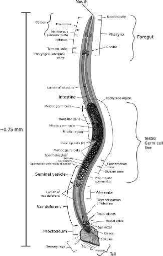
Anatomical drawing of a male C. elegans nematode with focus on the reproductive system. This free-living (not parasitic), colorless and transparent roundworm is approximately 1 mm in length, and lacks a respiratory and a circulatory system. (Wikimedia
...
- Adipose: Cell Types Composing the Tissue
- Adipose: Current Research on Isolation or Production of Therapeutic Cells
- Adipose: Development and Regeneration Potential
- Adipose: Existing or Potential Regenerative Medicine Strategies
- Adipose: Major Pathologies
- Adipose: Stem and Progenitor Cells in Adults
- Adipose: Tissue Function
- Adult Stem Cells: Overview
- Bladder: Cell Types Composing the Tissue
- Bladder: Current Research on Isolation or Production of Therapeutic Cells
- Bladder: Development and Regeneration Potential
- Bladder: Existing or Potential Regenerative Medicine Strategies
- Bladder: Major Pathologies
- Bladder: Stem and Progenitor Cells in Adults
- Blood Adult Stem Cell: Current Research on Isolation or Production of Therapeutic Cells
- Blood Adult Stem Cell: Development and Regeneration Potential
- Blood Adult Stem Cell: Existing or Potential Regenerative Medicine Strategies
- Blood Adult Stem Cell: Major Pathologies
- Blood Adult Stem Cell: Stem and Progenitor Cells in Adults
- Bone: Cell Types Composing the Tissue
- Bone: Current Research on Isolation or Production of Therapeutic Cells
- Bone: Development and Regeneration Potential
- Bone: Existing or Potential Regenerative Medicine Strategies
- Bone: Major Pathologies
- Bone: Stem and Progenitor Cells in Adults
- Breast: Cell Types Composing the Tissue
- Breast: Current Research on Isolation or Production of Therapeutic Cells
- Breast: Development and Regeneration Potential
- Breast: Existing or Potential Regenerative Medicine Strategies
- Breast: Major Pathologies
- Breast: Stem and Progenitor Cells in Adults
- Cartilage, Tendons, and Ligaments: Cell Types Composing the Tissue
- Cartilage, Tendons, and Ligaments: Current Research on Isolation or Production of Therapeutic Cells
- Cartilage, Tendons, and Ligaments: Development and Regeneration Potential
- Cartilage, Tendons, and Ligaments: Existing or Potential Regenerative Medicine Strategies
- Cartilage, Tendons, and Ligaments: Major Pathologies
- Cartilage, Tendons, and Ligaments: Stem and Progenitor Cells in Adults
- Dental: Current Research on Isolation or Production of Therapeutic Cells
- Dental: Development and Regeneration Potential
- Dental: Existing or Potential Regenerative Medicine Strategies
- Dental: Major Pathologies
- Dental: Stem and Progenitor Cells in Adults
- Eyes: Cell Types Composing the Tissue
- Eyes: Current Research on Isolation or Production of Therapeutic Cells
- Eyes: Development and Regeneration Potential
- Eyes: Existing or Potential Regenerative Medicine Strategies
- Eyes: Major Pathologies
- Eyes: Stem and Progenitor Cells in Adults
- Eyes: Tissue Function
- Gonads, Female: Cell Types Composing the Tissue
- Gonads, Female: Current Research on Isolation or Production of Therapeutic Cells
- Gonads, Female: Development and Regeneration Potential
- Gonads, Female: Major Pathologies
- Gonads, Female: Stem and Progenitor Cells in Adults
- Gonads, Male: Cell Types Composing the Tissue
- Gonads, Male: Current Research on Isolation or Production of Therapeutic Cells
- Gonads, Male: Development and Regeneration Potential
- Gonads, Male: Existing or Potential Regenerative Medicine Strategies
- Gonads, Male: Major Pathologies
- Gonads, Male: Stem and Progenitor Cells in Adults
- Graft Failure: Graft-Versus-Host Disease
- Gut: Current Research on Isolation or Production of Therapeutic Cells
- Gut: Development and Regeneration Potential
- Gut: Existing or Potential Regenerative Medicine Strategies
- Gut: Major Pathologies
- Gut: Stem and Progenitor Cells in Adults
- Heart: Cell Types Composing the Tissue
- Heart: Current Research on Isolation or Production of Therapeutic Cells
- Heart: Development and Regeneration Potential
- Heart: Existing or Potential Regenerative Medicine Strategies
- Heart: Major Pathologies
- Heart: Stem and Progenitor Cells in Adults
- Heart: Tissue Function
- Kidney: Cell Types Composing the Tissue
- Kidney: Current Research on Isolation or Production of Therapeutic Cells
- Kidney: Development and Regeneration Potential
- Kidney: Existing or Potential Regenerative Medicine Strategies
- Kidney: Major Pathologies
- Kidney: Stem and Progenitor Cells in Adults
- Liver: Cell Types Composing the Tissue
- Liver: Current Research on Isolation or Production of Therapeutic Cells
- Liver: Development and Regeneration Potential
- Liver: Existing or Potential Regenerative Medicine Strategies
- Liver: Major Pathologies
- Liver: Stem and Progenitor Cells in Adults
- Liver: Tissue Function
- Lung: Cell Types Composing the Tissue
- Lung: Current Research on Isolation or Production of Therapeutic Cells
- Lung: Development and Regeneration Potential
- Lung: Existing or Potential Regenerative Medicine Strategies
- Lung: Major Pathologies
- Lung: Stem and Progenitor Cells in Adults
- Lung: Tissue Function
- Mesenchymal: Cell Types Composing the Tissue
- Mesenchymal: Current Research on Isolation or Production of Therapeutic Cells
- Mesenchymal: Development and Regeneration Potential
- Mesenchymal: Existing or Potential Regenerative Medicine Strategies
- Mesenchymal: Major Pathologies
- Mesenchymal: Stem and Progenitor Cells in Adults
- Muscle: Cell Types Composing the Tissue
- Muscle: Current Research on Isolation or Production of Therapeutic Cells
- Muscle: Development and Regeneration Potential
- Muscle: Existing or Potential Regenerative Medicine Strategies
- Muscle: Major Pathologies
- Muscle: Stem and Progenitor Cells in Adults
- Neural: Cell Types Composing the Tissue
- Neural: Current Research on Isolation or Production of Therapeutic Cells
- Neural: Development and Regeneration Potential
- Neural: Existing or Potential Regenerative Medicine Strategies
- Neural: Major Pathologies
- Neural: Stem and Progenitor Cells in Adults
- Pancreas: Cell Types Composing the Tissue
- Pancreas: Current Research on Isolation or Production of Therapeutic Cells
- Pancreas: Development and Regeneration Potential
- Pancreas: Existing or Potential Regenerative Medicine Strategies
- Pancreas: Major Pathologies
- Pancreas: Stem and Progenitor Cells in Adults
- Pancreas: Tissue Function
- Skin: Cell Types Composing the Tissue
- Skin: Current Research on Isolation or Production of Therapeutic Cells
- Skin: Development and Regeneration Potential
- Skin: Existing or Potential Regenerative Medicine Strategies
- Skin: Major Pathologies
- Skin: Stem and Progenitor Cells in Adults
- Skin: Tissue Function
- Sweat Gland: Cell Types Composing the Tissue
- Sweat Gland: Current Research on Isolation or Production of Therapeutic Cells
- Sweat Gland: Development and Regeneration Potential
- Sweat Gland: Existing or Potential Regenerative Medicine Strategies
- Sweat Gland: Major Pathologies
- Sweat Gland: Stem and Progenitor Cells in Adults
- Vascular Stem Cell
- C. elegans Models to Study Stem Cells
- Drosophila Models to Study Stem Cells
- Mouse Models to Study Stem Cells
- Rat Models to Study Stem Cells
- Xenopus Models to Study Stem Cells
- Zebrafish Models to Study Stem Cells
- Brain Cancer
- Breast Cancer
- Cancer Stem Cells: Overview
- Colon Cancer
- Head and Neck Cancer
- Leukemia and Lymphoma Cancer Stem Cells
- Liver Cancer
- Lung Cancer
- Melanoma: Stem Cells
- Pancreatic Cancer
- Skin Cancer
- Clinical Trials, U.S.: AIDS-Related Conditions
- Clinical Trials, U.S.: Amyotrophic Lateral Sclerosis
- Clinical Trials, U.S.: Batten Disease
- Clinical Trials, U.S.: Blood Deficiencies
- Clinical Trials, U.S.: Crohn’s Disease
- Clinical Trials, U.S.: Diabetes
- Clinical Trials, U.S.: Eye Conditions
- Clinical Trials, U.S.: Graft Failure, Graft-Versus-Host Disease
- Clinical Trials, U.S.: Heart Disease
- Clinical Trials, U.S.: Hematological Cancers
- Clinical Trials, U.S.: Immunologic/Histiocytic Disorders
- Clinical Trials, U.S.: Kidney Disease
- Clinical Trials, U.S.: Multiple Sclerosis
- Clinical Trials, U.S.: Parkinson’s Disease
- Clinical Trials, U.S.: Peripheral Vascular Disease
- Clinical Trials, U.S.: Skin Transplants
- Clinical Trials, U.S.: Solid Tumors
- Clinical Trials, U.S.: Spinal Cord Injury
- Clinical Trials, U.S.: Stroke
- Clinical Trials, U.S.: Traumatic Brain Injury
- Clinical Trials Outside the United States
- Clinical Trials Outside the United States: Amyotrophic Lateral Sclerosis
- Clinical Trials Outside the United States: Cerebral Palsy
- Clinical Trials Outside the United States: Spinal Cord Injury
- Clinical Trials Outside the United States: Stroke
- Animal Cloning
- Reproductive and Therapeutic Cloning
- Australia
- Belgium
- Brazil
- China
- Denmark
- France
- Germany
- India
- Iran
- Italy
- Japan
- Korea
- Netherlands
- Norway
- Saudi Arabia
- Singapore
- Spain
- Sweden
- Switzerland
- Taiwan
- United Kingdom
- United States
- Chimera Formation, Ethics of
- Clinical Trials, Ethics of
- Clinical Trials (Adult Cells), Ethics of
- Cloning, Ethics of
- Egg Donation, Ethics of
- Genome Sequencing, Ethics of
- Germ Cell Modifications (Gene Therapy), Ethics of
- In Vitro Production of Germ Cells, Ethics of
- Moral Status of Embryos
- Unapproved Therapy, Ethics of
- Tissue Engineering (Scaffold)
- Tissue Printing
- Aastrom Biosciences, Inc.
- Advanced Cell Technology
- Athersys
- Beike Biotechnology
- California Stem Cell, Inc.
- Cellerant Therapeutics
- Cryo-Cell International
- Cryo-Save
- Cytori Therapeutics, Inc.
- ES Cell International
- Fate Therapeutics
- Geron Corporation
- International Stem Cell Corporation
- Lentigen Corporation
- NeoStem, Inc.
- Neuralstem, Inc.
- New Biotech: Overview
- Parcell Laboratories
- Pathfinder Cell Therapy
- Stem Cell Companies: Overview
- Stemagen
- Stematix
- TiGenix/Cellerix
- ViaCyte, Inc.
- Albert Einstein College of Medicine
- Autonomous University of Barcelona
- Autonomous University of Madrid
- Baylor University
- Boston Children’s Hospital
- California Institute for Regenerative Medicine
- Case Western Reserve University/Cleveland Clinic
- Columbia University
- Coriell Institute for Medical Research
- Duke University
- Genetics Policy Institute
- Harvard University
- Hebrew University of Jerusalem
- Howard Hughes Medical Institute
- Indiana University
- Johns Hopkins University
- Karolinska Institute
- Kyoto University
- Massachusetts General Hospital
- Massachusetts Institute of Technology
- Max Planck Society
- Mayo Clinic
- McMaster University
- Mount Sinai School of Medicine
- National Academy of Sciences
- Northwestern University
- Oregon Health & Science University
- Ottawa Hospital Research Institute
- Pasteur Institute
- Peking University
- Princeton University
- Reeve-Irvine Research Center
- Robarts Research Institute
- Rockefeller University
- Rutgers University
- Sanford-Burnham Medical Research Institute
- Scripps Research Institute
- Sloan Kettering Institute
- Stanford University
- Stowers Institute for Medical Research
- University of Bonn
- University of British Columbia
- University of California, Berkeley
- University of California, Davis
- University of California, Los Angeles
- University of California, San Diego
- University of California, San Francisco
- University of Cambridge
- University of Connecticut
- University of Heidelberg
- University of Melbourne
- University of Miami
- University of Michigan
- University of Milan
- University of Minnesota
- University of North Carolina at Chapel Hill
- University of Oxford
- University of Pittsburgh
- University of Southern California
- University of Strasbourg
- University of Texas Health Science Center at Houston
- University of Tokyo
- University of Toronto
- University of Washington/Fred Hutchinson Cancer Research Center
- University of Wisconsin–Madison
- University Pierre et Marie Curie, Paris
- Vanderbilt of University
- Wake Forest University
- Weill Cornell Medical College
- Weizmann Institute of Science
- Whitehead Institute for Biomedical Research
- Yale University
- Bioreactors
- Xenotransplantation
- Gene Patents
- International Society for Stem Cell Research Guidelines
- Pluripotent Stem Cell Patents
- Bone Marrow Transplants
- Direct Reprogramming of Adult Cells Into Other Cell Types
- Endothelial Cell Isolation
- Fluorescence-Activated Cell Sorting
- Genome Sequencing
- Human ES Cell Isolation
- In Vitro Fertilization
- In Vitro Stem Cell Study Assays
- In Vivo Stem Cell Study Assays
- Lineage Tracing
- Mouse ES Cell Isolation
- MRI Tracking
- Non-Human Primate Embryonic Stem Cells
- Nuclear Transfer, Altered
- Nuclear Transfer, Somatic Cell
- Parthogenesis
- Preimplantation Genetic Diagnosis
- Profiling Study Methods
- Stem Cell Epigenetic: Chromatin Modification
- Stem Cell Epigenetic: DNA Methylation
- Stem Cell Epigenetic: DNA Replication
- Stem Cell Expression Profiling
- Stem Cell Genetic Modification Study Method
- Stem Cell Markers
- Stem Cell Purification
- Tissue Culture Study Methods
- Viral Vectors: Adeonvirus
- Viral Vectors: Lentivirus
- Amniotic Fluid Cells
- Cord Blood Stem Cells
- Fetal Stem Cells
- Mesenchymal Stem Cells
- American Association for the Advancement of Science
- California Institute for Regenerative Medicine
- Canada: Stem Cell Network
- Christopher & Dana Reeve Foundation, The
- Danish Stem Cell Research
- EuroStemCell
- Howard Hughes Medical Institute
- International Society for Stem Cell Research
- International Stem Cell Forum
- Japan Human Cell Society
- Lasker Foundation
- Medical Research Council (UK)
- Michael J. Fox Foundation
- National Institutes of Health
- National Science Foundation
- National Stem Cell Bank
- New York Stem Cell Foundation
- Parkinson’s Disease Foundation
- Scotland: Stem Cell Research and Regenerative Medicine
- Stem Cell Genome Anatomy Projects
- Stem Cell Network North Rhine Westphalia
- Swiss Stem Cells Network
- UK National Stem Cell Network
- WiCell
- Alvarez-Buylla, Arturo
- Anversa, Piero
- Charo, Alta
- Frenette, Paul
- Fuchs, Elaine
- Goldman, Steven A.
- Hochedlinger, Konrad
- Langer, Robert
- Losordo, Douglas
- Martino, Gianvito
- McCulloch, Ernest
- Orkin, Stuart
- Pera, Martin
- Rafii, Shahin
- Rao, Mahendra
- Reynolds, Brent A., and Samuel Weiss
- Scadden, David
- Snyder, Evan
- Steindler, Dennis
- Studer, Lorenz
- Thomson, James
- Till, James
- van der Kooy, Derek
- Vescovi, Angelo
- Weissman, Irving
- Yamanaka, Shinya
- Embryonic Stem Cells, Methods to Produce
- iPS, Methods to Produce
- iPS Tissue Sources
- iPSC From Animal Cells
- Pluripotency Network
- Pluripotent Stem Cells, Embryonic
- Pluripotent Stem Cells, Epi
- Pluripotent Stem Cells, Germ
- Pluripotent Stem Cells, Teratoma
- Advocacy
- Congress: Votes and Amendments
- Do No Harm: The Coalition of Americans for Research Ethics
- National Right to Life Committee
- President’s Council on Bioethics
- Special Interest/Lobby Groups
- Stem Cells: Bush Ruling
- Buddhism
- Catholicism
- Christianity
- Judaism
- Muslim
- Alzheimer’s Disease
- Animal Cloning
- Autism
- Cord Blood Banking
- Cosmetic Surgery
- Drug Testing and Drug Development in Cell Culture
- Endothelial Blood Vessels
- ESC and iPSC Banking
- Fertility Treatment Creation of Germ Cells From Adult Cells
- Food From Stem Cells
- Gene Therapy: Hemoglobinopathies
- Graft Failure: Graft-Versus-Host Disease
- Hearing Disease
- Heart Disease
- Hematopoietic Transplantation: Cancer
- Hematopoietic Transplantation: Gene Therapy
- Immune Disorders
- In Utero Treatment
- Kidney Disease
- Lung Disease
- Pancreatic Islet Transplant
- Parkinson’s Disease
- Radiation Injury Treatment
- Retinal Stem Cells
- Schizophrenia
- Skin Graft
- Spinal Cord Injury
- Stem Cell Banking
- Transfusion Product: NK Cells
- Transfusion Product: Platelets
- Transfusion Product: Red Blood Cells
- Wound Repair
- Self-Renewal and Differentiation
- Stem Cell Aging
- Stem Cell Differentiation/Self-Organization
- Stem Cell DNA Repair
- Stem Cell Ground State
- Stem Cell Histocompatibility
- Stem Cell Niche
- Stem Cell Plasticity
- Stem Cell Potency
- Tissue Regeneration: Animals and Plants
- Tissue Regeneration: Humans
- Transit Amplifying Cells
- Alabama
- Alaska
- Arizona
- Arkansas
- California
- Colorado
- Connecticut
- Delaware
- Florida
- Georgia
- Hawai’i
- Idaho
- Illinois
- Indiana
- Iowa
- Kansas
- Kentucky
- Louisiana
- Maine
- Maryland
- Massachusetts
- Michigan
- Minnesota
- Mississippi
- Missouri
- Montana
- Nebraska
- Nevada
- New Hampshire
- New Jersey
- New Mexico
- New York
- North Carolina
- North Dakota
- Ohio
- Oklahoma
- Oregon
- Pennsylvania
- Rhode Island
- South Carolina
- South Dakota
- Tennessee
- Texas
- Utah
- Vermont
- Virginia
- Washington
- West Virginia
- Wisconsin
- Wyoming
- Loading...
Get a 30 day FREE TRIAL
-
 Watch videos from a variety of sources bringing classroom topics to life
 -
Read modern, diverse business cases
-
Explore hundreds of books and reference titles

Read next

More like this
Sage Recommends
We found other relevant content for you on other Sage platforms.
Have you created a personal profile? Login or create a profile so that you can save clips, playlists and searches