Entry
Reader's guide
Entries A-Z
Subject index
Neurosphere Cultures
THE ADULT CENTRAL nervous system has long been viewed as comprising tissue that is incapable of cell neogenesis and, particularly, lacking the ability to support production of new neuronal cells. Yet, discoveries made over the last two decades have radically altered this perspective. In fact, the discovery that some regions of the mature brain are the site of intense neurogenesis throughout life has changed our understanding of how the brain maintains its cy to architecture and functional integrity while, at the same time, possessing an inherent degree of plasticity and significant regeneration capacity.
The biological entity at the root of this neuro—genetic process is the neural stem cell. Although bearing somewhat distinct functional properties depending on their age and location, neural stem cells are involved in the production of new mature brain cells throughout life, including embryonic and fetal development. The in vitro approach that is most widely used to isolate and quantify these neural stem cells from the vast majority of the central nervous system tissues across species, higher primates and humans included, is the neurosphere technique that is described here. It is worth emphasizing how the neurosphere method also allows for the expansion of the neural stem cell population pool ex vivo while, at the same time, making it possible to measure critical stem cell features in the candidate neural cells, such as self—renewal, fate potential, and differentiation properties.
The Neurosphere Method
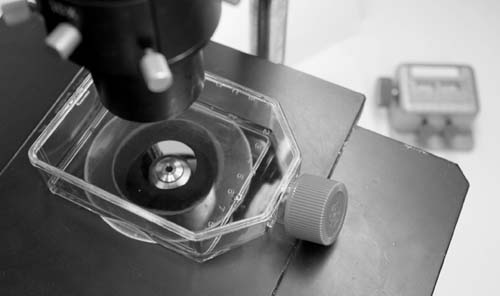
The neurosphere system can be applied to many different tissues, be they of adult, fetal, or embryonic origin, from virtually all mammalian species, including humans. In its most common application, donor tissue is predigested enzimatically and then mechanically dissociated to yield a single—cell suspension, which is then plated under quite stringent growth conditions. This procedure establishes a selective culture system in which most of the primary differentiated/differentiating central nervous system cells found in the primary tissue die out soon after plating, whereas the undifferentiated stem cells enter into a state of active proliferation.
The discovery that some regions of the mature brain are the site of intense neurogenesis throughout life has changed some of our understanding of the brain. The biological entity at the root of this process is the neural stem cell.
Four main conditions must absolutely be satisfied for the neural stem cells to become the prevalent cell type in these cultures: there must be low cell density (<5 × 10⁴ cells/cm2), there must be an absence of serum, and there must be the addition of the appropriate growth factors (i.e., EGF or FGF2), and there must be a plating substrate warranting loose cell adhesion (poly—L-lysine or poly—ornithine may be used). Under these conditions, cells from a freshly dissociated brain attach loosely to the substrate, with the majority (99 percent) rapidly dying out. At the same time, a tiny fraction of undifferentiated neural precursors, mostly neural stem cells, become hypertrophie, round up, and engage into active proliferation while adhering loosely to the culture vessel. The progeny of these proliferating precursors preferentially adhere to each other while dividing and, eventually, form spherical clusters that, because of their increasing mass, eventually lift off the substrate and float in suspension. These have been named neurospberes, from which comes the name of the technique.
...
- Biology
- Biotechnology, History of
- Cell Sorting
- Cells, Adult
- Cells, Amniotic
- Cells, Developing
- Cells, Embryonic
- Cells, Fetal
- Cells, Human
- Cells, Monkey
- Cells, Mouse (Embryonic)
- Cells, Neural
- Cells, Sources of
- Cells, Umbilical
- Cytogenetic Instability of Stem Cells
- Developmental Biology
- Differentiation, In Vitro and In Vivo
- Division Types (Symmetrical and Asymmetrical)
- Experimental Models
- Feeder/Feeder—Free Culture
- Gut Stem Cells
- Induced Pluripotent Stem Cells
- Lineages
- Mammary Stem Cells
- Markers of Sternness
- Methods of Growing Cells
- Microenvironment and Immune Issues
- Neuralstem
- Neurosphere Cultures
- Niche Self—Renewal
- Nuclear Reprogramming
- Parthogenesis
- Plant Stem Cells
- Prostate Tissue Stem Cells
- Renal Stem Cells
- Self—Renewal, Stem Cell
- Stem Cell Applications, Articular Cartilage
- Stem Cell Applications, Tendon and Ligament
- Stem—Like Cells, Human Brain
- Tissue Culture
- Transdifferentiation
- Clinical Trials
- Clinical Trials Outside U.S.: Amyotrophic Lateral Sclerosis
- Clinical Trials Outside U.S.: Avascular Necrosis
- Clinical Trials Outside U.S.: Severe Coronary Artery Disease
- Clinical Trials Outside U.S.: Spinal Cord Injury
- Clinical Trials Within U.S.: Batten Disease
- Clinical Trials Within U.S.: Blind Process
- Clinical Trials Within U.S.: Cancer
- Clinical Trials Within U.S.: Heart Disease
- Clinical Trials Within U.S.: Peripheral Vascular Disease
- Clinical Trials Within U.S.: Skin Transplants (Burns)
- Clinical Trials Within U.S.: Spinal Cord Injury
- Clinical Trials Within U.S.: Traumatic Brain Injury
- Clinical Trials Worldwide
- Countries
- Diseases
- Ethics
- History and Technology
- Birth Dating of Cells by Retrovirus
- Bone Marrow Transplants
- BrdU/Thymidine
- Fluorescence—Activated Cell Sorting
- Human Embryonic Stem Cells
- In Vitro Fertilization
- Mouse ES Cell Isolation
- MRI Tracking
- Non—Human Primate Embryonic Stem Cells
- Nuclear Transfer, Altered
- Nuclear Transfer, Somatic
- Parthogenesis
- Preimplantation Genetic Diagnosis
- Viral Vectors: Adeno—Associated Virus
- Viral Vectors: Adenovirus
- Viral Vectors: Lentivirus
- Industry
- Institutions
- Albert Einstein College of Medicine
- Baylor College of Medicine
- Bonn University
- Burnham Institute
- Caltech
- Cambridge University
- Case Western Reserve University/Cleveland Clinic
- Children's Hospital, Boston
- Columbia University
- Coriell Institute
- Duke University
- Genetics Policy Institute
- Harvard University
- Indiana University
- Johns Hopkins University
- Kyoto University
- Massachusetts General Hospital
- Massachusetts Institute of Technology
- Mayo Clinic
- McMaster University
- Mount Sinai School of Medicine
- National Academy of Science
- Northwestern University
- Oregon Health & Science University
- Ottawa Health Research Institute
- Oxford University
- Princeton University
- Reeve—Irvine Research Center
- Robarts Research Institute
- Rockefeller University
- Rutgers University
- Salk Institute
- Scripps Research Institute
- Sloan—Kettering Institute
- Stanford University
- Stowers Institute
- University of California, Berkeley
- University of California, Davis
- University of California, Los Angeles
- University of California, San Diego
- University of California, San Francisco
- University of Connecticut
- University of Georgia
- University of Miami
- University of Michigan
- University of Minnesota
- University of North Carolina, Chapel Hill
- University of Pittsburgh
- University of Southern California
- University of Texas Health Science Center at Houston
- University of Toronto
- University of Washington/Hutchinson Cancer Center
- University of Wisconsin, Madison
- Vanderbilt of University
- Wake Forest University
- Weill—Cornell Medical College
- Whitehead Institute
- Yale University
- Legal Issues
- Organizations
- American Association for the Advancement of Science
- Australian Stem Cell Centre
- California Institute for Regenerative Medicine
- Canadian Stem Cell Network
- China Stem Cell News
- Christopher Reeve Foundation
- Community of Stem Cell Scientists
- Danish Stem Cell Research Center
- East of England Stem Cell Network
- European Consortium for Stem Cell Research—EuroStemCell
- International Society for Stem Cell Research
- International Stem Cell Forum
- Japan Human Cell Society
- Lasker Foundation
- Medical Research Council UK Stem Cell Initiative
- Michael J. Fox Foundation
- National Institutes of Health
- National Stem Cell Bank
- Parkinson's Disease Foundation
- Scottish Stem Cell Network
- Stem Cell Genome Anatomy Projects
- Swiss Stem Cell Network
- UK National Stem Cell Network
- Wisconsin Alumni Research Foundation
- People
- Alvarez—Buylla, Arturo
- Anversa, Piero
- Charo, Robin Alta
- Eaves, Connie
- Eggan, Kevin
- Fuchs, Elaine
- Gage, Fred
- Gearhart, John
- Goldman, Steven A.
- Jaenisch, Rudolf
- Keller, Gordon
- Kriegstein, Arnold
- Lanza, Robert
- Losordo, Douglas
- Macklis, Jeffrey
- McKay, Ronald D. G.
- Melton, Doug
- Morrison, Sean
- Mummery, Christine
- Nottebohm, Fernando
- Okano, Hideyuki
- Orkin, Stuart
- Rao, Mahendra
- Smith, Austin
- Snyder, Evan
- Steindler, Dennis A.
- Studer, Lorenz P.
- Thomson, James
- Van der Kooy, Derek
- Verfaillie, Catherine
- Vescovi, Angelo
- Weissman, Irving
- Wilmut, Ian
- Politics
- Advocacy
- Coalition for the Advancement of Medical Research
- Congress: Votes and Amendments (Cloning/Embryos)
- Dickey Amendment
- Do No Harm: The Coalition of Americans for Research Ethics
- National Right to Life Committee
- President's Council on Bioethics
- Presidential Campaigns
- Reagan, Nancy
- Special Interest/Lobby Groups
- Stem Cells, Bush Ruling
- Religion
- States
- Alabama
- Arizona
- Arkansas
- California
- Colorado
- Connecticut
- Delaware
- Florida
- Georgia
- Hawaii
- Idaho
- Illinois
- Indiana
- Iowa
- Kansas
- Kentucky
- Louisiana
- Maine
- Maryland
- Massachusetts
- Michigan
- Minnesota
- Mississippi
- Missouri
- Montana
- Nebraska
- Nevada
- New Hampshire
- New Jersey
- New Mexico
- New York
- North Carolina
- North Dakota
- Ohio
- Oklahoma
- Oregon
- Pennsylvania
- Rhode Island
- South Carolina
- South Dakota
- Tennessee
- Texas
- Utah
- Vermont
- Virginia
- Washington
- West Virginia
- Wisconsin
- Wyoming
- Loading...
Get a 30 day FREE TRIAL
-
 Watch videos from a variety of sources bringing classroom topics to life
 -
Read modern, diverse business cases
-
Explore hundreds of books and reference titles

Read next

More like this
Sage Recommends
We found other relevant content for you on other Sage platforms.
Have you created a personal profile? Login or create a profile so that you can save clips, playlists and searches