
Entry
Reader's guide
Entries A-Z
Subject index
Slipped Disk
Spinal pain is a common reason for preventing athletes from participation in sports. The spine is composed of the cervical (neck), thoracic (upper back), and lumbar (low back) vertebrae. Approximately 20% of athletes will experience back pain during a 1-year period. Neck pain will occur in about 10% of athletes overall during this time period. Of course, this will vary from sport to sport. The cyclist will have a much higher prevalence of neck pain, while the gymnast will complain more of low back pain.
The sources of spinal pain are multiple and include the surrounding muscles, the ligaments, the facet joints, and the intervertebral disk. The disk accounts for up to 25% of spinal pain. However, there are a number of pathologic processes that are associated with the disk. It may degenerate with loss of fluid and height. Or the disk may rupture, which is called a herniated disk or, in lay terms, a “slipped disk.” To prevent and to properly rehabilitate an athlete with this injury, one must understand the biomechanics of both the sport and the individual athlete.
Anatomy
The spinal column descends from the skull to the pelvis with 7 cervical vertebrae, 12 thoracic vertebrae, and 5 lumbar vertebrae. The articulation of the adjacent-level vertebrae occurs via the intervertebral disk in the front and the facet joints in the back (Figure 1). The lumbar vertebrae end on the sacrum, which is a fused V-shaped vertebra that articulates with the pelvis to transfer forces from the trunk to the legs.
Figure 1 Lumbar Vertebrae (lateral view) and Single Vertebra (top view)
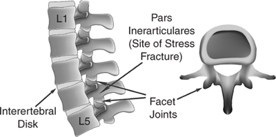
The disks become progressively larger from the cervical spine down through the lumbar spine. The disks provide shock absorption to the spine as well as motion. These disks are composed of three basic components: the annular ligaments on the outside (annulus), the colloidal gel (nucleus pulposus) on the inside, and the end plates of the vertebrae above and below. The annulus is composed of well-organized ligamentous sheaths (1020 layers; Figure 2). The nucleus pulposus is a hydrous gel with few cells and some inflammatory enzymes. The end plates are at the top and bottom of the vertebrae, where the disk attaches. Since there is no blood supply to the disk, the end plate provides the nutrition to the disk by way of hydrostatic pressure during motion. In the child and adolescent athletes, these end plates are composed of soft growth cartilage and are susceptible to injury.
The disks are round and crescent shaped, with the posterior curved away from the posterior spinal column. The one exception is the lowest disk at the lumbar-sacral juncture (L5-S1), which is round. In the adult, the disk has the weakest ligamentous constraints on the posterolateral side, where the nerve roots exit. As such, a rupture here will affect the nerve. Conversely, the child and young adolescent may rupture into the soft end plate growth cartilage.
Etiology
A slipped disk represents a herniation of the nucleus through a tear in the outer annular ligaments. When a small isolated tear occurs, the nucleus stays in place except for mild bulging. This annular tear will appear as an intense white line on some magnetic resonance imaging (MRI) scans. This is called a high-intensity zone (HIZ) and has been correlated with pain.
...
- Conditioning and Training
- Aerobic Endurance
- Body Composition (Body Mass Index)
- Burnout in Sports
- Cardiovascular and Respiratory Anatomy and Physiology: Responses to Exercise
- Circuit Training
- Conditioning
- Core Strength
- Cross-Training
- Detraining (Reversibility)
- Exercise Prescription
- Exercise Programs
- Fitness Testing
- Gender and Age Differences in Response to Training
- Home Exercise Equipment
- Immune System, Exercise and
- Interval Training/Fartlek
- Lean Body Weight Assessment
- Osteoporosis Prevention Through Exercise
- Overtraining
- Periodization
- Physiological Effects of Exercise on Cardiopulmonary System
- Plyometrics
- Principles of Training
- Resistance Training
- Speed, Agility, and Speed Endurance Development
- Static Stretching
- Strength Training for the Female Athlete
- Strength Training for the Young Athlete
- Stretching and Warming Up
- Target Heart Rate
- Temperature and Humidity, Effects on Exercise
- Women's Health, Effects of Exercise on
- Diagnosis and Treatment of Sports Injuries
- Acupuncture
- Ankle Support
- Arthroscopy
- Bracing
- Casting and Immobilization
- Complementary Treatment
- Crutches, How to Use
- Dual-Energy X-Ray Absorptiometry (DEXA)
- Electrical Stimulation
- Electromyography
- Extracorporeal Shock Wave Therapy
- Fieldside Assessment and Triage
- Joint Injection
- Joints, Magnetic Resonance Imaging of
- Nonsteroidal Anti-Inflammatory Drugs (NSAIDs)
- Operating Room Equipment and Environment
- Orthotics
- Pain Management in Sports Medicine
- Pharmacology and Exercise
- Physical Examination and History
- Preparticipation Cardiovascular Screening
- Presports Physical Examination
- PRICE/MICE
- Taping
- Ultrasound
- Musculoskeletal Examination Techniques
- Diet and Nutrition
- Calcium in the Athlete's Diet
- Carbohydrates in the Athlete's Diet
- Dietary Supplements and Vitamins
- Fat in the Athlete's Diet
- Nutrition and Hydration
- Postgame Meal
- Pregame Meal
- Protein in the Athlete's Diet
- Salt in the Athlete's Diet
- Sports Drinks
- Vegetarianism and Exercise
- Weight Gain for Sports
- Weight Loss for Sports
- Doping and Performance Enhancement
- Alcohol and Exercise
- Anabolic Steroids
- Beta Agonists
- Beta Blockers
- Blood Transfusion
- Caffeine and Exercise
- Cannabinoids
- Creatine
- Diuretics
- Doping and Performance Enhancement: A New Definition
- Doping and Performance Enhancement: Historical Overview
- Doping and Performance Enhancement: Olympic Games From 2004 to 2008
- Erythropoietin
- Growth Hormone
- Narcotic Analgesics
- Peptide Hormones
- Performance Enhancement, Doping, Therapeutic Use Exemptions
- Stimulants
- World Anti-Doping Agency
- Exercise Physiology, Biomechanics, and Kinesiology
- Injuries and Disorders
- Altitude Sickness
- Athlete's Heart Syndrome
- Bursitis
- Catastrophic Injuries
- Clavicle (Collarbone) Fracture
- Cold Injuries and Hypothermia
- Contusions (Bruises)
- Cramping
- Cyst, Ganglion
- Dehydration
- Epicondylitis
- Exertional Compartment Syndrome, Chronic
- Fractures
- Handlebar Palsy
- Headache, Exercise-Induced
- Hyponatremia, Exercise-Associated
- Lightning Injuries
- Muscle Cramps
- Organ Injuries
- Pectoralis Strain
- Referred Pain
- Rhinitis, Exercise-Induced
- Side Stitch
- Sports Injuries, Acute
- Sports Injuries, Overuse
- Strains, Muscle
- Stress Fractures
- Sudden Cardiac Death
- Tendinitis, Tendinosis
- Tendinopathy
- Trigger Finger
- Trunk Injuries
- Abdomen
- Ankle
- Back and Lumbar Spine
- Back Injuries, Surgery for
- Cervical and Thoracic Fractures and Traumatic Instability
- Cervical Brachialgia
- Intervertebral Disk Disease
- Klippel-Feil Syndrome
- Lower Back Contusion
- Lower Back Injuries and Low Back Pain
- Lower Back Muscle Strain and Ligament Sprain
- Rhomboid Muscle Strain and Spasm
- Slipped Disk
- Spinal Cord Injury
- Spondylolysis and Spondylolisthesis
- Cervical and Thoracic Spine
- Cervical and Thoracic Disk Disease
- Cervical and Thoracic Fractures and Traumatic Instability
- Cervical and Thoracic Spine Injuries
- Cervical Brachialgia
- Cervical Disk Degeneration
- Cervical Facet Syndrome
- Costosternal Syndrome (Costochondritis)
- Intervertebral Disk Disease
- Os Odontoideum
- Scheuermann Kyphosis
- Scoliosis
- Chest Wall
- Elbow and Forearm
- Biceps Tendinitis
- Biceps Tendon Rupture
- Elbow and Forearm Injuries
- Elbow and Forearm Injuries, Surgery for
- Elbow Bursitis
- Elbow Dislocations
- Elbow Fractures
- Elbow Sprain
- Elbow, Osteochondritis Dissecans of the
- Forearm Fracture
- Little League Elbow
- Medial Apophysitis of the Elbow
- Medial Epicondyle Avulsion Fractures of the Elbow
- Olecranon Stress Injury
- Panner Disease
- Posterior Impingement Syndrome
- Posterolateral Rotatory Instability
- Ulnar Neuropathy
- Foot
- Arch Pain
- Athlete's Foot
- Black Nail
- Blisters
- Bruised Foot
- Bunions
- Calluses
- Corns
- Foot and Ankle Injuries, Surgery for
- Foot Fracture
- Foot Injuries
- Foot Stress Fracture
- Hammertoe
- Ingrown Toenail
- Metatarsalgia
- Morton Neuroma
- Overpronating Foot
- Oversupinating Foot
- Plantar Fasciitis and Heel Spurs
- Retrocalcaneal Bursitis
- Sesamoiditis
- Sever Disease
- Turf Toe
- Warts (Verrucae)
- Hand and Finger
- Head and Neck
- Cervical Nerve Stretch Syndrome
- Concussion
- Craniofacial Injuries
- Dental Injuries
- Detached Retina
- Diffuse Axonal Injury
- Ear Infection, Outer (Otitis Externa)
- Ear Injuries
- Epidural Hematoma
- Eye Injuries
- Facial Injuries
- Head Injuries
- Intracerebral Hematoma
- Intracranial Hemorrhage
- Malignant Brain Edema Syndrome
- Neck and Upper Back Injuries
- Neck Spasm
- Nose Injuries
- Punch Drunk Syndrome
- Skull Fracture
- Subarachnoid Hemorrhage
- Subdural Hematoma
- Torticollis, Acute
- Hip, Pelvis, and Groin
- Avascular Necrosis of the Femoral Head
- Avulsion Fractures
- Femoral Neck Stress Fracture
- Femoroacetabular Impingement
- Genitourinary Injuries
- Gluteal Strain
- Groin Pain
- Groin Strain
- Groin Tendinitis
- Groin/Inguinal Hernia
- Hip Contusion
- Hip Dislocation
- Hip Flexor Strain
- Hip Flexor Tendinitis
- Hip Fracture
- Hip Stress Fracture
- Hip, Pelvis, and Groin Injuries
- Hip, Pelvis, and Groin Injuries, Surgery for
- Osteitis Pubis
- Pelvic Avulsion Fractures
- Pelvic Bursitis
- Pelvic Fracture
- Pelvic Stress Fracture
- Perineal Numbness and Erectile Dysfunction
- Piriformis Syndrome
- Sacroiliac Pain
- Sciatica
- Slipped Capital Femoral Epiphysis
- Snapping Hip Syndrome
- Tailbone (Coccyx) Injuries
- Knee
- Anterior Cruciate Ligament Tear
- Articular and Meniscal Cartilage Regeneration and Repair
- Chondromalacia Patella
- Cyst, Baker
- Discoid Meniscus
- Extensor Mechanism Injury
- Iliotibial Band Syndrome
- Juvenile Osteochondritis Dissecans of the Knee
- Knee Bursitis
- Knee Injuries
- Knee Injuries, Surgery for
- Knee Ligament Sprain, Medial and Lateral Collateral Ligaments
- Knee Plica
- Knee, Osteochondritis Dissecans of the
- Kneecap, Subluxating
- Meniscus Injuries
- Osgood-Schlatter Disease
- Patellar Dislocation
- Patellar Tendinitis
- Patellofemoral Pain Syndrome
- Posterior Cruciate Ligament Injuries
- Quadriceps Tendinitis
- Tibial Tubercle Avulsion Fracture
- Lower Leg
- Shoulder
- Frozen Shoulder
- Glenoid Labrum Tear
- Little League Shoulder
- Rotator Cuff Tears, Partial
- Rotator Cuff Tendinopathy
- Shoulder Arthritis
- Shoulder Bursitis
- Shoulder Dislocation
- Shoulder Impingement Syndrome
- Shoulder Injuries
- Shoulder Injuries, Surgery for
- Shoulder Instability
- Shoulder Subluxation
- Superior Labrum From Anterior to Posterior (SLAP) Lesions
- Skin
- Abrasions and Lacerations
- Allergic Contact Dermatitis
- Angioedema and Anaphylaxis
- Athlete's Nodules
- Dermatology in Sports
- Friction Injuries to the Skin
- Frostbite and Frost Nip
- Fungal Skin Infections and Parasitic Infestations
- Insect Bites and Stings
- Irritant Contact Dermatitis
- Jock Itch
- Jogger's Nipples
- Pressure Injuries to the Skin
- Prickly Heat
- Puncture Wounds
- Skin Conditions in Wrestlers
- Skin Disorders, Metabolic
- Skin Infections, Bacterial
- Skin Infections, Viral
- Skin Infestations, Parasitic
- Sunburn
- Sunburn and Skin Cancers
- Toenail Fungus
- Urticaria and Pruritus
- Warts (Verrucae)
- Thigh
- Thorax
- Wrist
- Injury Prevention
- Medical Conditions Affecting Sports Participation
- Allergies
- Altitude Sickness
- Anaphylaxis, Exercise-Induced
- Anemia
- Anorexia Nervosa
- Arthritis
- Asthma
- Asthma, Exercise-Induced
- Atlantoaxial Instability
- Bleeding Disorders
- Bruised Ribs
- Burnout in Sports
- Cholinergic Urticaria
- Complex Regional Pain Syndrome
- Congenital Heart Disease
- Dehydration
- Detached Retina
- Diabetes in the Young Athlete
- Diabetes Mellitus
- Diarrhea
- Exercise and Heart Murmurs
- Exercise and Mitral Valve Prolapse
- Fever
- Functionally One-Eyed Athlete
- Gastrointestinal Problems
- Heat Illness
- Hepatitis
- Hypertension (High Blood Pressure)
- Infectious Diseases in Sports Medicine
- Ingrown Toenail
- Jet Lag
- Liver Conditions, Hepatitis, Hepatomegaly
- Marfan Syndrome
- Neurologic Disorders Affecting Sports Participation
- Obesity
- Respiratory Conditions
- Seizure Disorder in Sports
- Sickle Cell Disease
- Sinusitis in Athletes
- Skin Disorders Affecting Sports Participation
- Anatomical Abnormalities
- Infectious Diseases
- Blood-Borne Infections
- Ear Infection, Outer (Otitis Externa)
- Epstein-Barr Virus, Infectious Mononucleosis, and Splenomegaly
- Fungal Skin Infections and Parasitic Infestations
- Hepatitis
- HIV and the Athlete
- Infectious Diseases in Sports Medicine
- Jock Itch
- Methicillin-Resistant Staphylococcus Aureus Infections
- Pulmonary and Cardiac Infections in Athletes
- Skin Conditions in Wrestlers
- Skin Infections, Bacterial
- Skin Infections, Viral
- Skin Infestations, Parasitic
- Toenail Fungus
- Rehabilitation and Physical Therapy
- Special Populations
- Specialties and Occupations in Sports Medicine
- Athletic Trainers
- Dietitian/Sports Nutritionist
- Emergency Medicine and Sports
- Exercise Physiologist
- Family Doctor
- Group Fitness Instructor
- Manual Medicine
- Orthopedist in Sports Medicine, Role of
- Physiatry and Sports Medicine
- Physical and Occupational Therapist
- Podiatric Sports Medicine
- Sport and Exercise Psychology
- Sports Biomechanist
- Sports Massage Therapist
- Team Physician
- Sport Psychology
- Anger and Violence in Sports
- Arousal and Athletic Performance
- Attention Focus in Sports
- Biofeedback
- Bulimia Nervosa
- Burnout in Sports
- Exercise Addiction/Overactivity Disorders
- Hypnosis and Sport Performance
- Imagery and Visualization
- Leadership in Sports
- Mental Health Benefits of Sports and Exercise
- Motivation
- Overtraining
- Personality and Exercise
- Psychological Aspects of Injury and Rehabilitation
- Psychological Assessment in Sports
- Sport and Exercise Psychology
- Sports Socialization
- Team and Group Dynamics in Sports
- Sports and Society
- Air Pollution, Effects on Exercise and Sports
- Anger and Violence in Sports
- Benefits of Exercise and Sports
- Diversity in Sports
- Doping and Performance Enhancement: A New Definition
- Doping and Performance Enhancement: Historical Overview
- Doping and Performance Enhancement: Olympic Games From 2004 to 2008
- Epidemiology of Sports Injuries
- Legal Aspects of Sports Medicine
- Protective Equipment in Sports
- Sports Injuries, Overuse
- Team and Group Dynamics in Sports
- Title IX, Education Amendments of 1972
- World Anti-Doping Agency
- Sports and Sports Medicine
- Air Pollution, Effects on Exercise and Sports
- Anatomy and Sports Medicine
- Benefits of Exercise and Sports
- Circadian Rhythms and Exercise
- Diversity in Sports
- Emergency Medicine and Sports
- Epidemiology of Sports Injuries
- Exercise and Disease Prevention
- Future Directions in Sports Medicine
- History of Sports Medicine
- Immune System, Exercise and
- Physical Examination and History
- Seasonal Rhythms and Exercise
- Shift Work and Exercise
- Skill Acquisition in Sports
- Sleep and Exercise
- Sleep Loss, Effects on Athletic Performance
- Sports Injuries, Surgery for
- Travel Medicine and the International Athlete
- Women's Health, Effects of Exercise on
- Organizations
- Issues for Practitioners
- Sports-Specific Injuries
- Aerobic Dance, Injuries in
- Archery, Injuries in
- Badminton, Injuries in
- Baseball, Injuries in
- Basketball, Injuries in
- Biking, Injuries in
- Boxing, Injuries in
- Cheerleading, Injuries in
- Cricket, Injuries in
- Dance Injuries and Dance Medicine
- Extreme Sports, Injuries in
- Field Hockey, Injuries in
- Figure Skating, Injuries in
- Football, Injuries in
- Golf, Injuries in
- Gymnastics, Injuries in
- Horse Riding, Injuries in
- Ice Hockey, Injuries in
- Karate, Injuries in
- Marathons, Injuries in
- Mixed Martial Arts, Injuries in
- Mountain Bike Racing, Injuries in
- Outdoor Athlete
- Racquetball and Squash, Injuries in
- Rowing, Injuries in
- Rugby Union, Injuries in
- Running Injuries
- Sailing and Yacht Racing, Injuries in
- SCUBA Diving, Injuries in
- Skiing, Injuries in
- Snowboarding, Injuries in
- Soccer, Injuries in
- Speed Skating, Injuries in
- Surfing, Injuries in
- Swimming, Injuries in
- Tennis and Racquet Sports, Injuries in
- Tennis Elbow
- Triathlons, Injuries in
- Ultimate Frisbee, Injuries in
- Volleyball, Injuries in
- Weight Lifting, Injuries in
- Windsurfing, Injuries in
- Wrestling, Injuries in
- Women and Sports
- Amenorrhea in Athletes
- Dysmenorrhea
- Eating Disorders
- Exercise During Pregnancy and Postpartum
- Female Athlete
- Female Athlete Triad
- Gender and Age Differences in Response to Training
- Menstrual Cycle and Sports Performance
- Menstrual Irregularities
- Osteoporosis Prevention Through Exercise
- Title IX, Education Amendments of 1972
- Transsexual Athletes
- Women's Health, Effects of Exercise on
- Loading...
Get a 30 day FREE TRIAL
-
 Watch videos from a variety of sources bringing classroom topics to life
 -
Read modern, diverse business cases
-
Explore hundreds of books and reference titles

Read next

More like this
Sage Recommends
We found other relevant content for you on other Sage platforms.
Have you created a personal profile? Login or create a profile so that you can save clips, playlists and searches