Entry
Reader's guide
Entries A-Z
Subject index
Glaucoma
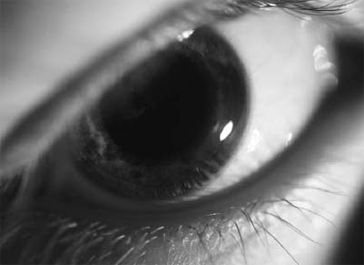
Glaucoma is a disease characterized by elevated intraocular pressure, optic nerve damage, and subsequent impaired vision. Glaucoma is one of the leading causes of adult blindness in the United States and worldwide. Risk factors include increased age, African-American race, and family history in a first-degree relative. The main types of glaucoma include primary open angle glaucoma, angle closure glaucoma, and congenital glaucoma. Primary open angle glaucoma is the most common form of glaucoma and occurs in 0.5 to 2.1 percent of the population over the age of 40. It is caused by a resistance to flow of aqueous humor. Aqueous humor is produced in the ciliary body and drained by the trabecular meshwork. Dysfunction of the trabecular meshwork leads to a disruption in the normal flow of aqueous humor. This results in an increase in the pressure of the eye and damage to the optic nerve. The increased pressure causes a decrease in the blood supply to the nerve and thus damage to the nerve. Over time, due to the damage to the nerve, patients will gradually lose peripheral vision. In advanced cases, the vision loss can lead to absolute blindness.
Angle closure glaucoma results from the peripheral iris, or colored part of the eye, blocking the trabecular meshwork. The angle closure leads to a rise of the in traocular pressure from its normal level (10 to 21 mm Hg) to 30 mm Hg or more. This results in an increase in aqueous fluid inside the eye causing damage to the optic nerve. Prodromal symptoms occur as transitory attacks during which time patients may experience symptoms of decreased vision, eye pain, halos around lights, headache, nausea, and vomiting.
Glaucoma is one of the leading causes of adult blindness in the United States and worldwide.
Another important type of glaucoma is congenital or infantile glaucoma. Congenital glaucoma has an incidence of 1 in 10,000 births with an increased incidence in males. The exact cause is unknown but appears related to a maldevelopment of the aqueous humor drainage system. Signs and symptoms include enlarged cornea (buphthalmos), photophobia, and tearing. Prompt surgical intervention offers the best method of controlling the intraocular pressure and long-term preservation of vision.
For the adult patient, the first signs of glaucoma include gradual loss of peripheral vision. Patients with angle closure glaucoma may present with pain and colored halos, but with primary open-angle glaucoma, patients may be asymptomatic until late in the disease. Glaucoma screening involves a complete ophthalmic history and examination, including evaluation of intraocular pressures, optic nerve, visual fields, and gonioscopy (evaluation of the drainage system of the eye). Intraocular pressure in most individuals ranges between 10 and 21 mmHg, with an average of approximately 16 mmHg. Patients with pressures above 21 are suspicious for glaucoma. Intraocular pressures can fluctuate throughout the day, so pressure levels cannot be the only screening tool for glaucoma.
During examination, the optic nerve is also observed. The optic nerve is examined by comparing the cup to disk ratio. A central depression exists in the optic nerve called the cup. An increase in the size of the cup relative to the rest of the nerve signifies glaucomatous damage. Physical examination should also include gonioscopy. Gonioscopy consists of a special lens placed on the eye that allows visualization of the trabecular meshwork, peripheral iris, cornea, and presence of angle closure. For patients with glaucoma, management includes intraocular pressure checks every three to six months, visual field examination every six to 12 months, gonioscopy and optic nerve evaluation yearly.
...
- Children's Health
- Adolescent Development
- Adolescent Health
- Adoption Medicine
- Alcohol and Youth
- Asthma in Children
- Birth Defects
- Breech Birth
- Child Abuse
- Child Behavior Disorders
- Child Dental Health
- Child Development
- Child Mental Health
- Child Safety
- Childhood Cancers
- Childhood Immunization
- Diabetes Type I (Juvenile Diabetes)
- Dysmorphology
- Exercise for Children
- Failure to Thrive
- Fetal Alcohol Syndrome
- Hearing Problems in Children
- Infant and Newborn Care
- Infant and Toddler Development
- Infant and Toddler Health
- Juvenile Rheumatoid Arthritis
- Neonatologist
- Neonatology
- Pediatrics
- Premature Babies
- Prenatal Care
- Rickets
- Smoking and Youth
- Spina Bifida
- Sudden Infant Death Syndrome
- Tanner Stages
- Countries: Africa
- Algeria
- Angola
- Benin
- Botswana
- Brunei Darussalam
- Burkina Faso
- Burundi
- Côte d'Ivoire
- Cameroon
- Cape Verde
- Central African Republic
- Chad
- Comoros
- Congo
- Congo, Democratic Republic
- Djibouti
- Egypt
- Equatorial Guinea
- Eritrea
- Ethiopia
- Gabon
- Gambia
- Ghana
- Guinea
- Guinea-Bissau
- Kenya
- Lesotho
- Liberia
- Madagascar
- Malawi
- Mali
- Mauritania
- Mauritius
- Morocco
- Mozambique
- Namibia
- Niger
- Nigeria
- Rwanda
- São Tomé and Principe
- Senegal
- Sierra Leone
- Somalia
- South Africa
- Sudan
- Swaziland
- Tanzania
- Togo
- Tunisia
- Uganda
- Zambia
- Zimbabwe
- Countries: Americas
- Antigua and Barbuda
- Argentina
- Bahamas
- Barbados
- Belize
- Bolivia
- Brazil
- Canada
- Chile
- Colombia
- Costa Rica
- Cuba
- Dominica
- Dominican Republic
- Ecuador
- El Salvador
- Grenada
- Guatemala
- Guyana
- Haiti
- Honduras
- Jamaica
- Mexico
- Nicaragua
- Panama
- Paraguay
- Peru
- Saint Kitts and Nevis
- Saint Lucia
- Saint Vincent and the Grenadines
- Suriname
- Trinidad and Tobago
- United States Statistics
- Uruguay
- Venezuela
- Countries: Asia
- Afghanistan
- Azerbaijan
- Bahrain
- Bangladesh
- Bhutan
- Cambodia
- China
- East Timor
- Georgia
- India
- Indonesia
- Iran
- Iraq
- Israel
- Japan
- Jordan
- Kazakhstan
- Korea, North
- Korea, South
- Kuwait
- Kyrgyzstan
- Laos
- Lebanon
- Malaysia
- Maldives
- Moldova
- Mongolia
- Myanmar
- Nepal
- Oman
- Pakistan
- Palestine
- Philippines
- Qatar
- Russia
- Saudi Arabia
- Seychelles
- Singapore
- Sri Lanka
- Syria
- Tajikistan
- Thailand
- Turkey
- Turkmenistan
- Ukraine
- United Arab Emirates
- Uzbekistan
- Vietnam
- Yemen
- Countries: Europe
- Albania
- Andorra
- Armenia
- Austria
- Belarus
- Belgium
- Bosnia and Herzegovina
- Bulgaria
- Croatia
- Cyprus
- Czech Republic
- Denmark
- Estonia
- Finland
- France
- Germany
- Greece
- Hungary
- Iceland
- Ireland
- Italy
- Latvia
- Liechtenstein
- Lithuania
- Luxembourg
- Macedonia FYROM
- Malta
- Monaco
- Netherlands
- Norway
- Poland
- Portugal
- Romania
- San Marino
- Serbia and Montenegro
- Slovakia
- Slovenia
- Spain
- Sweden
- Switzerland
- United Kingdom
- Countries: Pacific
- Diseases, Cancers
- Adrenocortical Carcinoma
- Anal Cancer
- Bile Duct Cancer
- Bladder Cancer
- Bone Cancer
- Brain Cancer
- Breast Cancer
- Cancer (General)
- Carcinoid Tumors
- Carcinoma of Unknown Primary
- Colorectal Cancer
- Esophageal Cancer
- Extragonadal Germ Cell Tumor
- Eye Cancer
- Gallbladder Cancer
- Head and Neck Cancer
- Hodgkin's Lymphoma
- Kidney Cancer
- Leukemia
- Liver Cancer
- Lung Cancer
- Malignant Mesothelioma
- Multiple Myeloma
- Neuroblastoma
- Non-Hodgkin Lymphoma
- Oncologist
- Oncology
- Oral Cancer
- Osteonecrosis
- Ovarian Cancer
- Pancreatic Cancer
- Penile Cancer
- Pheochromocytoma
- Pituitary Tumor
- Prostate Cancer
- Skin Cancer
- Small Intestine Cancer
- Soft Tissue Sarcoma
- Stomach Cancer
- Testicular Cancer
- Thymus Cancer
- Thyroid Cancer
- Uterine Cancer
- Vaginal Cancer
- Vulvar Cancer
- Diseases, Localized
- Acid Reflux
- Acne
- Acoustic Neuroma
- Age-Related Macular Degeneration
- Alzheimer's Disease
- Amblyopia
- Anal/Rectal Diseases
- Aneurysms
- Angina
- Aphasia
- Appendicitis
- Arrhythmia
- Arteriosclerosis
- Asbestos/Asbestosis
- Asthma
- Back Injuries
- Back Pain
- Bell's Palsy
- Birthmark
- Bladder Diseases
- Blepharitis
- Blepharospasm
- Blindness
- Bone Diseases
- Bone Marrow Diseases
- Brain Diseases
- Brain Injuries
- Brain Malformations
- Breast Diseases
- Breathing Problems
- Bronchitis
- Carpal Tunnel Syndrome
- Cataract
- Celiac Disease
- Cellulitis
- Chlamydia Infections
- Chronic Obstructive Pulmonary Disease (COPD)
- Cirrhosis
- Cleft Lip and Palate
- Colon Polyps
- Colonic Diseases (General)
- Concussion
- Congenital Heart Disease
- Conjunctivitis
- Connective Tissue Disorders
- Constipation
- Cornea and Corneal Disease
- Coronary Disease
- Deafness
- Dental Health
- Dermatitis
- Diabetic Eye Problems
- Diabetic Foot
- Diabetic Kidney Problems
- Diabetic Nerve Problems
- Diabetic Teeth and Gum Problems
- Diarrhea
- Digestive Diseases (General)
- Diverticulosis and Diverticulitis
- Dysphagia
- Ear Disorders
- Ear Infections
- Elbow Injuries and Disorders
- Emphysema
- Esophagus Disorders
- Eye Diseases (General)
- Facial Injuries and Disorders
- Foot Health
- Foot Injuries and Disorders
- Fractures
- Gallbladder and Bile Duct Diseases
- Gastroesophageal Reflux/Hiatal Hernia
- Gastrointestinal Bleeding
- Genetic Brain Disorders
- Genital Herpes
- Glaucoma
- Glomerular Diseases
- Goiter
- Gonorrhea
- Gout and Pseudogout
- Gum Disease
- Hair Diseases and Hair Loss
- Hand Injuries and Disorders
- Head and Brain Injuries
- Head and Brain Malformations
- Head Lice
- Headache and Migraine
- Heart Attack
- Heart Diseases (General)
- Heart Valve Diseases
- Hemorrhoids
- Hepatitis
- Hepatitis C
- Hernia
- Hip Injuries and Disorders
- Huntington's Disease
- Hydrocephalus
- Impetigo
- Incontinence
- Inflammatory Bowel Disease
- Intestinal Parasites
- Irritable Bowel Syndrome
- Kidney Diseases (General)
- Kidney Failure and Dialysis
- Kidney Stones
- Knee Injuries and Disorders
- Leg Injuries and Disorders
- Liver Diseases (General)
- Low Vision
- Meniere's Disease
- Mouth Disorders
- Neck Disorders and Injuries
- Neural Tube Defects
- Nose Disorders
- Onchocerciasis
- Paget's Disease of Bone
- Pancreatic Diseases
- Peptic Ulcer
- Pneumonia
- Polycystic Kidney Disease
- Pulmonary Embolism
- Pulmonary Fibrosis
- Raynaud's Disease
- Respiratory Diseases (General)
- Retinal Disorders
- Rheumatic Heart Disease
- Rosacea
- Salivary Gland Disorders
- Scoliosis
- Severe Acute Respiratory Syndrome (SARS)
- Shoulder Injuries and Disorders
- Sinusitis
- Skin Diseases (General)
- Skin Pigmentation Disorders
- Spinal Cord Diseases
- Spinal Cord Injuries
- Spinal Muscular Atrophy
- Spinal Stenosis
- Spleen Diseases
- Stomach Disorders
- Taste and Smell Disorders
- Temporomandibular Joint Dysfunction
- Thoracic Outlet Syndrome
- Throat Disorders
- Thyroid Diseases
- Tinnitus
- Trachoma
- Tuberculosis
- Ulcers
- Urinary Tract Infections
- Usher Syndrome
- Vitiligo
- Voice Disorders
- Wrist/Arm Injuries and Disorders
- Diseases, Systemic
- E. Coli Infections
- Acquired Mutation
- Addison's Disease
- AIDS
- AIDS and Infections
- AIDS-Related Malignancies
- Alcoholism
- Allergy
- Anemia
- Anorexia Nervosa
- Arthritis
- Autoimmune Diseases (General)
- Bacterial Infections
- Beriberi
- Bird Flu
- Bleeding Disorders
- Botulism
- Bulimia
- Bursitis
- Cerebral Palsy
- Chagas Disease
- Chickenpox
- Cholera
- Chronic Fatigue Syndrome
- Coma
- Common Cold
- Cystic Fibrosis
- Degenerative Nerve Diseases
- Dengue
- Developmental Disabilities
- Diabetes
- Diabetes Type II
- Diphtheria
- Disabilities (General)
- Dizziness and Vertigo
- Down Syndrome
- Dystonia
- Eating Disorders
- Edema
- Ehlers-Danlos Syndrome
- Endocrine Diseases (General)
- Epilepsy
- Fainting
- Fever
- Filariasis/Elephantiasis
- Food Allergy
- Foodborne Diseases
- Fungal Infections
- Gaucher's Disease
- Genetic Disorders
- Germline Mutation
- Giardia Infections
- Growth Disorders
- Guillain-Barre Syndrome
- Hemorrhagic Fever
- Herpes Simplex
- High Blood Pressure
- Hyperactivity
- Hypoglycemia
- Hypothermia
- Immune System and Disorders
- Infectious Diseases (General)
- Influenza
- Klinefelter's Syndrome
- Kwarshiorkor
- Lactose Intolerance
- Lead Poisoning
- Legionnaire's Disease
- Leishmaniasis
- Leprosy
- Leukodystrophies
- Lou Gehrig's Disease
- Low Blood Pressure
- Lupus
- Lyme Disease
- Lymphatic Diseases
- Malaria
- Marasmus
- Marfan Syndrome
- Measles
- Meningitis
- Metabolic Disorders
- Missense Mutation
- Movement Disorders
- Multiple Chemical Sensitivity
- Multiple Sclerosis
- Mumps
- Muscle Disorders
- Muscular Dystrophy
- Myasthenia Gravis
- Myositis
- Nausea and Vomiting
- Neurofibromatosis
- Neurologic Diseases (General)
- Neuromuscular Disorders
- Nonsense Mutation
- Osteoarthritis
- Osteogenesis Imperfecta
- Pain
- Paralysis
- Parasitic Diseases
- Parathyroid Disorders
- Parkinson's Disease
- Peripheral Nerve Disorders
- Phenylketonuria
- Pituitary Disorders
- Point Mutation
- Poisoning
- Polio and Post-Polio Syndrome
- Polymyalgia Rheumatica
- Porphyria
- Prader-Willi Syndrome
- Psoriasis
- Rabies
- Reflex Sympathetic Dystrophy
- Rett Syndrome
- Reye Syndrome
- Rheumatoid Arthritis
- Rubella
- Salmonella Infections
- Sarcoidosis
- Scleroderma
- Seizures
- Sexually Transmitted Diseases
- Shingles (Herpes Zoster)
- Shistosomiasis
- Sickle Cell Anemia
- Sjogren's Syndrome
- Smallpox
- Somatic Mutation
- Staphylococcal Infections
- Streptococcal Infections
- Stress
- Stroke
- Tay-Sachs Disease
- Tetanus
- Tourette Syndrome
- Transient Ischemic Attack
- Tremor
- Tuberous Sclerosis
- Turner's Syndrome
- Typhoid
- Vasculitis
- Viral Infections
- Whooping Cough
- Wilson's Disease
- Drugs and Drug Companies
- Adult Immunization
- Amphetamines
- Anabolic Steroids
- Antibiotics
- Drug and Medical Device Safety
- Generic Drug
- Immunization/Vaccination
- Inhalants
- Maintenance Medication
- Me-Too Drug
- Over-the-Counter (OTC) Drug
- Pharmaceutical Industry (Worldwide)
- Pharmacist
- Pharmacoepidemiology
- Pharmacogenetics
- Pharmacogenomics
- Pharmacologist
- Pharmacology
- Pharmacopeia/Pharmacopoeia
- Pharmacy
- Placebo
- Prescription Drugs Abuse
- United States Pharmacopeia and National Formulary (USP-NF)
- Health Sciences
- Acquired Immunity
- Active Immunity
- Activities of Daily Living (ADLs)
- Aerospace Medicine
- African American Health
- AIDS, Living with
- Air Pollution
- Alcohol Consumption
- Allele
- Allopathy
- Alpha Error
- Alternative Medicine
- Alzheimer's Caregivers
- Amputees
- Anthrax
- Antioxidants
- Artificial Limbs
- Asian American Health
- Background Radiation
- Bacteriology
- Base Excision Repair
- Base Pair
- Base Sequence
- Beta Error
- Biochemistry
- Biodefense
- Bioinformatics
- Biological and Chemical Weapons
- Biomarker
- Biosafety
- Bioterrorism
- Birth Cohort
- Birth Rate
- Bisexual
- Body Cell Mass
- Body Mass Index
- Body Surface Area
- Bone Health
- Bone Mass Density
- Bone Mineral Density
- Brown Fat
- Burns
- Caffeine
- Calcium
- Cancer—Coping with Cancer
- Cardiologist
- Cardiology
- Caregivers
- Carotenoid
- Cascade
- Centenarian
- Cervical Spine
- Chemokine
- Chemokinesis
- Chinese Medicine, Traditional
- Cholesterol
- Chromosome
- Chronology of Global Health
- Circadian
- Clone
- Club Drugs
- Cocaine Abuse
- Coefficient of Inbreeding
- Complex Humanitarian Emergencies
- Conventional Medicine
- Craniology
- Crossing Over
- Crude Mortality Rate
- Cytogenetics
- Cytokine
- Daily Reference Values (DRVs)
- Date Rape
- Death and Dying
- Death Rate
- Demographic Transition
- Diesel Exhaust
- Dieting
- Disasters and Emergency Preparedness
- Disease and Poverty
- Disease Prevention
- Dizygotic Twin
- DNA
- DNA Repair
- Drinking Water
- Drug Abuse
- Dyslexia
- Ecogenetics
- Ectoparasites
- Elder Abuse
- Electromagnetic Fields
- Electrophysiology
- Empiric Risk
- Endemic
- Endocrinology
- Engram
- Environmental Health
- Environmental Medicine
- Environmental Tobacco Smoke (ETS)
- Environmental Toxicology
- Epidemic
- Epidemiologist
- Epidemiology
- Epigenetics
- Evidence-Based Medicine
- Exercise for Seniors
- Exercise/Physical Fitness
- Eye Care
- Falls
- False Negative
- False Positive
- First American Medical School
- Flea Bites
- Fluoride
- Folic Acid
- Food Contamination/Poisoning
- Food Safety
- Forensic Medicine
- Fraternal Twins
- Gastroenterologist
- Gastroenterology
- Gay Gene
- Gene Pool
- Genetic Code
- Genetics
- Genomic Library
- Genotype
- Geriatrics
- Gerontology
- Global Health Ethics
- Hair Dye
- Haploid
- Hardy-Weinberg Law
- Healthcare, Africa
- Healthcare, Asia and Oceania
- Healthcare, Europe
- Healthcare, South America
- Healthcare, U.S. and Canada
- Heart Diseases—Prevention
- Heat Index
- Hematologist
- Hematology
- Hemizygous
- Hemochromatosis
- Herbal Medicine
- Herbal Remedy
- Herbalism
- Herbalist
- Heroin Abuse
- Heterosexual
- Heterozygote
- Hispanic American Health
- Histology
- Homeopathy
- Homicide
- Homosexual
- Homozygote
- Hormones
- Household Poisons
- Hygiene
- Immunologist
- Immunology
- In Situ
- In Vitro
- In Vivo
- Inbreeding
- Incidence
- Indoor Air Pollution
- Infertility
- Innate Immunity
- Inpatient
- Institutional Review Board (IRB)
- Internal Medicine
- Internist
- Iridology
- Knockout
- Lean Body Mass
- Learning Disorders
- Lesbian
- Locus
- Malariologist
- Malariology
- Marijuana Abuse
- Medical College Admissions Test (MCAT)
- Medical Entomology
- Medical Geography
- Medical Helminthology
- Medical Jurisprudence
- Medical Physics
- Medical Tourism
- Memory
- Mercury
- Meta-analysis
- Methamphetamine Abuse
- Methylation
- Mismatch Repair
- Molds
- Monozygotic Twin
- Mortality
- Mosquito Bites
- Native American Health
- Naturopathy
- Nephrologist
- Nephrology
- Neuroendocrinology
- Neurologist
- Neurology
- Neuropsychologist
- Neuroradiologist
- Neuroradiology
- Neuroscience
- Neuroscientist
- Noise
- Nuclear Medicine
- Nutrition
- Nutritionist
- Obesity
- Occupational Health
- Occupational Injuries
- Occupational Medicine
- Ophthalmologist
- Ophthalmology
- Optometrist
- Oral Surgeon
- Organ Donation
- Orthomolecular Medicine
- Orthopedics
- Orthopedist
- Osteopathy
- Otolaryngologist
- Otolaryngology
- Otology
- Outpatient
- Ozone
- Passive Immunity
- Pasteurization
- Pathologist
- Pathology
- Pathophysiology
- Perinatologist
- Perinatology
- Pesticides
- Phenotype
- Phrenology
- Phylogenetics
- Physiatrist
- Physical Therapist
- Physiology
- Podiatrist
- Pollution
- Polymerase Chain Reaction (PCR)
- Polymorphism
- Prevalence
- Preventive Care
- Primary Care
- Proctology
- Programmed Cell Death
- Pulmonology
- Radiation Exposure
- Radiologist
- Radiology
- Radon
- Rare Diseases
- Refractive Errors
- Refugee Health
- Rehabilitation
- Reproductive Health (General)
- Roentgenology
- Safety (General)
- Satellite DNA
- Saturated Fat
- Sleep Disorders
- Smokeless Tobacco
- Smoking
- Smoking Cessation
- Snellen's Chart
- Speech & Communication Disorders
- Spider Bites
- Sports Injuries
- Sprains and Strains
- Sun Exposure
- Sun Protection Factor (SPF)
- Telepathology
- Third World
- Tick Bites
- Toxicology
- Trans Fat
- Travel Medicine
- Traveler's Health
- Tropical Medicine
- Tumor Registry
- Ultraviolet Radiation
- United States Medical Licensing Examination (USMLE)
- United States Statistics
- Universal Donor
- Unsaturated Fat
- Urological Surgeon
- Urologist
- Virology
- Vitamin A Deficiency
- Vitamin and Mineral Supplements
- West Nile Virus
- Men's Health
- Mental Health
- Anxiety Disorders
- Attention Deficit Disorder
- Attention Deficit Disorder with Hyperactivity
- Autism
- Bereavement
- Dementia
- Depression
- Manic-Depressive Illness
- Mental Health
- Obsessive-Compulsive Disorder
- Psychiatrist
- Psychiatry
- Psychoimmunology
- Psychologist
- Psychology
- Psychoneuroimmunology
- Psychooncology
- Psychotherapy
- Schizophrenia
- Suicide
- Organizations and Associations
- Adult Congenital Heart Association (ACHA)
- Alzheimer's Association
- Alzheimer's Disease Education and Referral Center (ADEAR)
- American Academy of Addiction Psychiatry (AAAP)
- American Academy of Family Physicians (AAFP)
- American Academy of Nurse Practitioners (AANP)
- American Academy of Ophthalmology (AAO)
- American Academy of Orthopedic Surgeons (AAOS)
- American Academy of Pediatrics (AAP)
- American Association for Cancer Research (AACR)
- American Association for Health Education (AAHE)
- American Association of Orthodontists (AAO)
- American Cancer Society (ACS)
- American College Health Association (ACHA)
- American College of Epidemiology (ACE)
- American College of Nurse-Midwives (ACNM)
- American College of Obstetricians and Gynecologists (ACOG)
- American College of Occupational and Environmental Medicine (ACOEM)
- American College of Physicians (ACP)
- American College of Preventive Medicine (ACPM)
- American College of Sports Medicine (ACSM)
- American College of Surgeons (ACS)
- American Council for Fitness and Nutrition (ACFN)
- American Dental Association (ADA)
- American Dental Hygienists' Association (ADHA)
- American Diabetes Association (ADA)
- American Fertility Association (AFA)
- American Geriatrics Society (AGS)
- American Health Care Association (AHCA)
- American Heart Association (AHA)
- American Lung Association
- American Medical Association (AMA)
- American Medical Women's Association (AMWA)
- American Nurses Association (ANA)
- American Obesity Association (AOA)
- American Osteopathic Association
- American Pharmaceutical Association (APhA)
- American Podiatric Medical Association (APMA)
- American Pregnancy Association (APA)
- American Psychological Association (APA)
- American Public Health Association (APHA)
- American Red Cross
- American Social Health Association (ASHA)
- American Society for Reproductive Medicine (ASRM)
- American Society of Clinical Oncology (ASCO)
- American Society of Clinical Pathologists (ASCP)
- American Society of Human Genetics (ASHG)
- American Society on Aging (ASA)
- American Urological Association (AUA)
- Association for International Cancer Research (AICR)
- Association of Maternal and Child Health Programs (AMCHP)
- Association of Schools of Public Health (ASPH)
- Association of Women's Health, Obstetric and Neonatal Nurses (AWHONN)
- European Association for Cancer Research (EACR)
- European Association for the Study of Obesity (EASO)
- Fondation Jean Dausset (CEPH)
- Human Genome Organisation (HUGO)
- Institute for Children's Environmental Health (ICEH)
- Institute of Medicine (IOM)
- International AIDS Vaccine Initiative (IAVI)
- International Center for Equal Healthcare Access (ICEHA)
- International Classification of Diseases (ICD)
- International Clinical Epidemiology Network (INCLEN)
- International Committee of the Red Cross (ICRC)
- International Council of AIDS Service Organizations (ICASO)
- International Epidemiological Association (IEA)
- International Federation of Red Cross and Red Crescent Societies (IFRC)
- International Genetic Epidemiology Society (IGES)
- International Red Cross and Red Crescent Movement (RCRC)
- International Society for Environmental Epidemiology (ISEE)
- International Society for Pharmacoepidemiology (ISPE)
- International Society of Geographical and Epidemiological Ophthalmology (ISGEO)
- International Women's Health Coalition (IWHC)
- Médicins Sans Frontières
- MedicAlert
- National Asian Women's Health Organization (NAWHO)
- National Association of Health Data Organizations (NAHDO)
- National Association of People with AIDS (NAPWA)
- National Breast Cancer Coalition (NBCC)
- National Coalition for Cancer Survivorship (NCCS)
- National Environmental Health Association (NEHA)
- National Mental Health Association (NMHA)
- National Network for Immunization Information (NNii)
- National Women's Health Organization (NWHO)
- North American Association for the Study of Obesity (NAASO)
- Pasteur Institute
- School Nutrition Association (SNA)
- Society for Healthcare Epidemiology of America (SHEA)
- Society for Nutrition Education (SNE)
- Society for Public Health Education (SOPHE)
- Voluntary Euthanasia Society (VES)
- People
- Abse, Dannie
- Ames, Bruce N.
- Avery, Oswald Theodore
- Axelrod, Julius
- Beals, Rodney K.
- Beijerinck, Martinus W.
- Bell, Charles
- Blackwell, Elizabeth
- Bross, Irwin D.J.
- Brown, Louise
- Brown, Michael Stuart
- Calabresi, Paul
- Casals-Ariet, Jordi
- Chekhov, Anton
- Chen, Zhong Wei
- Crick, Francis
- Da Vinci, Leonardo
- Darwin, Charles
- Ehrlich, Paul
- Farmer, Paul
- Fredrickson, Donald
- Gage, Phineas
- Galton, Sir Francis
- Gibbon, John H., Jr.
- Hardy, James D.
- Hounsfield, Godfrey
- Hughlings Jackson, John
- Kübler-Ross, Elisabeth
- Kelman, Charles D.
- Kirklin, John W.
- La Montagne, John
- Lauterbur, Paul C.
- Lederberg, Joshua
- Lewis, Edward B.
- Mansfield, Peter
- Marx, Gertie F.
- McClintock, Barbara
- Mead, Margaret
- Moscati, St. Joseph
- Nirenberg, Marshall W.
- Osler, Sir William
- Parkinson, James
- Pasteur, Louis
- Pauling, Linus
- Ramsay Hunt, James
- Rodbell, Martin
- Roentgen, Wilhelm
- Sabin, Albert
- Sabin, Florence R.
- Sachs, Jeffrey
- Schweitzer, Albert
- Soper, Fred L.
- Stewart, Alice
- Stickler, Gunnar B.
- Thomas, Lewis
- Thorn, George W.
- Varco, Richard L.
- Warshaw, Joseph
- Watson, James
- Wilkins, Lawson
- Zoll, Paul M.
- Procedures and Therapies
- Acupuncture
- Allograft
- Angioplasty
- Biofeedback
- Biotherapy
- Blood/Blood Transfusion
- Cancer Alternative Therapy
- Cancer Chemotherapy
- Cancer Radiation Therapy
- Cardioversion
- Chemoprevention
- Chemoradiotherapy
- Chemotherapy
- Chiropractic
- Cryopreservation
- Cryosurgery
- Diagnostic Imaging
- Diagnostic Tests
- Dialysis
- Enzyme-Linked Immunosorbent Assay (ELISA)
- Exercise Treadmill Test
- Gel Electrophoresis
- Gene Array Analysis
- Gene Mapping
- Gene Silencing
- Gene Transfer
- Genes and Gene Therapy
- Genetic Testing/Counseling
- Genetic Transformation
- Genomic Imprinting
- Heart Bypass Surgery
- Heart Transplantation
- Immunosuppression
- Immunotherapy
- Kidney Transplantation
- Laboratory Tests
- Liver Transplantation
- Lung Transplantation
- Microsurgery
- Oral Rehydration Therapy
- Organ Transplantation
- Pancreas Transplantation
- Stem Cells/Stem Cell Transplantation
- Surgery
- Telesurgery
- Research
- Society and Health
- Administration for Children and Families (ACF)
- Administration on Aging (AoA)
- Agency for Healthcare Research and Quality (AHRQ)
- Agency for Toxic Substances and Disease Registry (ATSDR)
- Center for Food Safety and Applied Nutrition (CFSAN)
- Centers for Disease Control and Prevention (CDC)
- Centers for Medicare and Medicaid Services (CMS)
- Department of Energy (DOE)
- Department of Health and Human Services (HHS)
- Drug Enforcement Administration (DEA)
- Employment Retirement Income Security Act (ERISA)
- Environmental Protection Agency (EPA)
- European Food Safety Authority (EFSA)
- European Public Health Alliance (EPHA)
- European Public Health Association (EUPHA)
- Federal Emergency Management Agency (FEMA)
- Fee-for-Service
- Food and Agriculture Organization of the United Nations (FAO)
- Food and Drug Administration (FDA)
- Global Health Council
- Health Insurance Portability and Accountability Act (HIPAA)
- Health Maintenance Organization (HMO)
- Health Resources and Services Administration (HRSA)
- Indian Health Service (IHS)
- Insurance
- International Agency for Research on Cancer (IARC)
- International Health Ministries Office (IHMO)
- Joint FAO/WHO Expert Committee on Food Additives (JECFA)
- Joint United Nations Programme in HIV/AIDS (UNAIDS)
- Managed Care
- Medicaid
- Medical Research Council
- Medicare
- Medigap Policy
- MEDLINE
- Mine Safety and Health Administration (MSHA)
- National Cancer Institute (NCI)
- National Center for Chronic Disease Prevention and Health Promotion (NCCDPHP)
- National Center for Complementary and Alternative Medicine (NCCAM)
- National Center for Environmental Health (NCEH)
- National Center for Health Marketing (NCHM)
- National Center for Health Statistics (NCHS)
- National Center for HIV, STD, and TB Prevention (NCHSTP)
- National Center for Infectious Diseases (NCID)
- National Center for Injury Prevention and Control (NCIPC)
- National Center for Public Health Informatics (NCPHI)
- National Center on Birth Defects and Developmental Disabilities (NCBDDD)
- National Cholesterol Education Program (NCEP)
- National Eye Institute (NEI)
- National Heart, Lung, and Blood Institute (NHLBI)
- National Human Genome Research Institute (NHGRI)
- National Immunization Program (NIP)
- National Institute of Allergy and Infectious Diseases (NIAID)
- National Institute of Arthritis and Musculoskeletal and Skin Diseases (NIAMS)
- National Institute of Biomedical Imaging and Bioengineering (NIBIB)
- National Institute of Children's Health (NICHD)
- National Institute of Diabetes and Digestive and Kidney Diseases (NIDDK)
- National Institute of Environmental Health Sciences (NIEHS)
- National Institute of Mental Health (NIMH)
- National Institute of Neurological Disorders and Stroke (NINDS)
- National Institute of Nursing Research (NINR)
- National Institute of Occupational Safety and Health (NIOSH)
- National Institute on Aging (NIA)
- National Institute on Alcohol Abuse and Alcoholism (NIAAA)
- National Institute on Deafness and Other Communication Disorders (NIDCD)
- National Institute on Drug Abuse (NIDA)
- National Institutes of Health (NIH)
- National Library of Medicine (NLM)
- National Program of Cancer Registries (NPCR)
- Occupational Safety and Health Administration (OSHA)
- Pan American Health Organization (PAHO)
- Point of Service (POS)
- Preferred Provider Organization (PPO)
- Public Health
- Substance Abuse and Mental Health Services Administration (SAMHSA)
- United Nations Children's Fund (UNICEF)
- United Network for Organ Sharing (UNOS)
- United States Public Health Service
- World Health Organization (WHO)
- Women's Health
- Abortion
- AIDS
- AIDS and Pregnancy
- Birth Control/Contraception
- Bisexual
- Breast Cancer
- Breast Feeding
- Breast Implants/Breast Reconstruction
- Cervical Cancer
- Climacteric
- Diabetes and Pregnancy
- Doula
- Estrogen Replacement Therapy (ERT)
- Female Circumcision
- Gynecologist
- Gynecology
- Heterosexual
- High Risk Pregnancy
- Hormone Replacement Therapy
- Hormones
- Infertility
- Lesbian
- Menopause
- Menstruation and Premenstrual Syndrome
- Midwife
- Obstetric Fistula
- Obstetrician/Gynecologist
- Obstetrics
- Osteoporosis
- Ovarian Cancer
- Postpartum Depression
- Preeclampsia
- Pregnancy
- Pregnancy and Substance Abuse
- Pregnancy Loss
- Reproductive Health (General)
- Smoking and Pregnancy
- Teenage Pregnancy
- Urinary Tract Infections
- Uterine Cancer
- Uterine Diseases
- Vaginal Cancer
- Vaginal Diseases
- Vulvar Cancer
- Women's Health (General)
- Loading...
Get a 30 day FREE TRIAL
-
 Watch videos from a variety of sources bringing classroom topics to life
 -
Read modern, diverse business cases
-
Explore hundreds of books and reference titles

Read next

More like this
Sage Recommends
We found other relevant content for you on other Sage platforms.
Have you created a personal profile? Login or create a profile so that you can save clips, playlists and searches