Entry
Reader's guide
Entries A-Z
Subject index
Brain Research
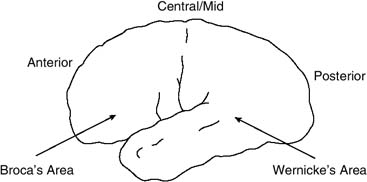
The study of language and the brain has a long history, beginning with reports in the 1800s of language impairment aphasia, meaning a loss or impairment of the power to use or comprehend words, usually resulting from brain damage. These early cases demonstrated that damage to different areas of the left hemisphere of the brain produced different types of language deficit (see Figure 1). Lesions to the temporal lobe, specifically Wernicke's area, compromise the ability to understand language and the ability to speak clearly. Lesions to the frontal cortex, Broca's area, prevent a person from producing speech. For example, a person with a lesion in this area has the ability to understand language, but words are not properly formed, and speech is slow and slurred.
Initially, these correlations were established postmortem. However, with the advent of the computed tomography (CT) scan in the 1970s, it became possible to examine brain damage in living patients and to explore changes in brain structure as a patient recovered.
During the past 15 years, there has been a marked increase in the availability of brain imaging techniques to language scientists. These techniques have made it possible, for the first time, to study brain activity correlated with language learning and processing in unimpaired subjects and in some cases to examine this activity within a short time frame. Two of these techniques have been put to relatively greater use than others, and these will be the focus of this entry.
With respect to bilingualism, imaging studies permit systematic exploration of ideas stemming from earlier reports; these indicated that seizure disorders, stroke, and other injuries, along with localized electrical stimulation during brain surgery, can affect one language and leave others unaffected in a bilingual or multilingual individual. Further, imaging studies open a new window into the perennial questions of how the age of learning a second language (L2) and degree of achieved fluency influence the way the L2 is represented in the person's mind and brain. One must be cautious when interpreting results that explore the localization of language in bilin-guals, since there is some variation (particularly in localization and lateralization) even across monolin-guals. However, mounting evidence shows that second languages learned relatively late, or not learned fluently, are physically represented in somewhat different regions of the brain than the first language (LI) is and are processed in a different time frame.
Figure 1 Left Hemisphere of Brain, With Approximate Locations of Broca's Area and Wernicke's Area
Magnetic Resonance Imaging (MRI)
The first imaging technique discussed here is functional magnetic resonance imaging. The general technique of magnetic resonance imaging (MRI) surrounds a region of the body with a high-intensity magnetic field and beams radio waves through it, creating high-resolution topographic images of tissue. Functional MRI measures changes in the metabolic activity (including blood flow and oxy-genation) of specific portions of the brain during a specified activity, such as language use. Functional MRI is the technique of choice when the critical issue is localization of function within the brain; it can discriminate between small regions less than a millimeter apart in the brain. The temporal resolution of functional MRI is not as great as with event-related potentials (ERP), described below, restricting the range of questions that can be asked about the time course of information retrieval and processing. Functional MRI can produce a new image of the brain and the changes in it once every second.
...
- Family, Communities, and Society
- Accommodation Theory, Second-Language
- Americanization and its Critics
- Attitudes toward Language Diversity
- Benefits of Bilingualism and Heritage Languages
- Bilingual Education in the Press
- Easy and Difficult Languages
- English in the World
- English-Only Organizations
- Heritage Languages in Families
- Hidden Curriculum
- Hispanic Population Growth
- Home/School Relations
- Immigration and Language Policy
- Language Brokering
- Language Loyalty
- Language Restrictionism
- Nationality-Culture Myth
- One Person-One Language (OPOL)
- Peer Pressure and Language Learning
- Raising Bilingual Children
- Spanish Loan Words in U.S. English
- Spanish, Decline in use
- Spanish, The Second National Language
- Transnational Students
- Views of Language Difference
- History
- Americanization and its Critics
- Boarding Schools and Native Languages
- Defense Language Institute
- Early Bilingual Programs, 1960s
- Early Immigrants and English Language Learning
- Equity Struggles and Educational Reform
- German Language Education
- German Language in U.S. History
- Languages in Colonial Schools, Eastern
- Languages in Colonial Schools, Western
- Latino Civil Rights Movement
- National Education Association Tucson Symposium
- Nationalization of Languages
- Navajo Code Talkers
- President's Commission on Foreign Language and International Studies
- Puerto Rico, School Language Policies
- Southeast Asian Refugees
- St. Lambert Immersion Study
- Vietnamese Immigration
- Instructional Designs
- Additive and Subtractive Programs
- Biculturalism
- Bilingual Charter Schools
- Bilingual Special Education
- Costs of Bilingual Education
- Deaf Bilingual Education
- Designation and Redesignation of English Language Learners
- Dual-Language Programs
- English as a Second Language Approaches
- English Immersion
- English or Content Instruction
- Gifted and Talented Bilinguals
- Heritage Language Education
- Indigenous Language Revitalization
- Indigenous Languages as Second Languages
- Literacy and Biliteracy
- Multicultural Education
- Newcomer Programs
- Oyster Bilingual School
- P.S. 25, New York City's First Bilingual School
- Phonics in Bilingual Education
- Program Goals, Purpose of
- Program Quality Indicators
- Pull-Out ESL Instruction
- Sheltered Instruction Observation Protocol
- Spanish, Proactive Maintenance
- Specially Designed Academic Instruction in English
- Transitional Bilingual Education Programs
- Whole Language
- Languages and Linguistics
- Accents and Their Meaning
- Affective Filter
- Baby Talk
- BICS/CALP Theory
- Bilingualism Stages
- Chinese in the United States
- Chinese Language Study, Prospects
- Code Switching
- Cognates, True and False
- Compound and Coordinate Bilingualism
- Comprehensible Input
- Container Theory of Language
- Continua of Biliteracy
- Critical Languages for the United States
- Critical Period Hypothesis
- Discourse Analysis
- Ebonics
- English, First World Language
- First-Language Acquisition
- Indigenous Languages, Current Status
- Indo-European Languages
- Interlanguage
- Japanese Language in Hawai'i
- Language Acquisition Device
- Language Defined
- Language Dominance
- Language Persistence
- Language Registers
- Language Revival and Renewal
- Language Shift and Language Loss
- Language Socialization
- Language Socialization of Indigenous Children
- Learning a Language, Best Age
- Linguistics, an Overview
- Measuring Language Proficiency
- Metalinguistic Awareness
- Modern Languages in Schools and Colleges
- Monitor Model
- Native English Speakers Redefined
- Phonology, Morphology, and Syntax
- Pragmatics
- Second-Language Acquisition
- Semilingualism
- Skills Transfer Theory
- Social Bilingualism
- Spanglish
- Threshold Hypothesis
- Underlying Linguistic Proficiencies
- World Englishes
- People and Organizations
- Alatis, James E.
- Andersson, Theodore
- Baker, Colin
- Bennett, William J.
- Bernal, Joe J.
- Bourne, Randolph S.
- Cárdenas, José A.
- Castro Feinberg, Rosa
- Center for Applied Linguistics, Initial Focus
- Center for Applied Linguistics, Recent Focus
- Chavez, Linda
- Christian, Donna
- Collier, Virginia P.
- Crawford, James
- Cummins, James
- De Avila, Edward
- Epstein, Noel
- Escamilla, Kathy
- Escobedo, Deborah
- Fernández, Ricardo
- Fishman, Joshua A.
- Gómez, Joel
- Gómez, Severo
- García, Eugene E.
- González, Henry B.
- González, Josué M.
- Guerrero, Adalberto
- Hakuta, Kenji
- Haugen, Einar
- Hayakawa, S. I.
- Hogan, Timothy M.
- Hornberger, Nancy
- Kloss, Heinz
- Krashen, Stephen D.
- LaFontaine, Hernán
- Lyons, James J.
- Moll, Luis
- Multicultural Education, Training, and Advocacy (META)
- National Association for Bilingual Education
- National Clearinghouse for Bilingual Education
- Nieto, Sonia
- Office for Civil Rights, U.S. Department of Education
- Office of Bilingual Education and Minority Languages Affairs
- Ogbu, John
- Oyama, Henry
- Pérez-Hogan, Carmen
- Peña, Álbar Antonio
- Porter, Rosalie Pedalino
- Rodríguez, Armando
- Rodríguez, Richard
- Roos, Peter D.
- Roybal, Edward R.
- Ruiz, Richard
- Saville-Troike, Muriel
- Seidner, María M.
- Simon, Paul M.
- Spolsky, Bernard
- Stanford Working Group
- Tanton, John H.
- TESOL, Inc.
- Troike, Rudolph C, Jr.
- Truán, Carlos
- Trueba, Enrique (Henry)
- Unz, Ron
- Urquides, María
- Valdés, Guadalupe
- Wong Fillmore, Lily
- Yarborough, Ralph
- Zamora, Gloria L.
- Zelasko, Nancy
- Policy Evolution
- Castañeda Three-Part Test
- Flores v. State of Arizona
- Lau v. Nichols, Enforcement Documents
- Lau v. Nichols, San Francisco Unified School District's Response
- Lau v. Nichols, the Ruling
- Méndez v. Westminster
- Affirmative Steps to English
- Amendment 31 (Colorado)
- Aspira Consent Decree
- Bilingual Education as Language Policy
- Canadian and U.S. Language Policies
- Chacón-Moscone Legislation
- Civil Rights Act of 1964
- English for the Children Campaign
- Equal Educational Opportunity Act of 1974
- Exit Criteria for English Language Learner Programs
- Federal Court Decisions and Legislation
- High-Stakes Testing
- Home Language Survey
- Immigration and Language Policy
- Improving America's Schools Act of 1994
- Labeling Bilingual Education Clients: LESA, LEP, and ELL
- Language Education Policy in Global Perspective
- Language Policy and Social Control
- Language Rights in Education
- Maintenance Policy Denied
- National Defense Education Act of 1958
- National Literacy Panel
- Native American Languages, Legal Support for
- No Child Left Behind Act of 2001, Testing Requirements
- No Child Left Behind Act of 2001, Title I
- No Child Left Behind Act of 2001, Title III
- Official English Legislation, Favored
- Official English Legislation, Position of English Teachers on
- Official Language Designation
- Paradox of Bilingualism
- Proposition 203 (Arizona)
- Proposition 203 (Arizona), Impact of
- Proposition 227 (California)
- Proposition 227 (California), Impact of
- Question 2 (Massachusetts)
- Texas Legislation (HB 103 and SB 121)
- Title VII, Elementary and Secondary Education Act, 1967 Senate Hearings
- Title VII, Elementary and Secondary Education Act, Key Historical Marker
- Title VII, Elementary and Secondary Education Act, Subsequent Amendments
- Title VII, Elementary and Secondary Education Act, Text (Appendix B)
- Transitional Bilingual Education Model Questioned
- U.S. Bilingual Education Viewed from Abroad
- U.S. Commission on Civil Rights Report
- Undocumented Students' Rights
- Voter Initiatives in Education
- Related Social Sciences
- Acculturation
- Affective Dimension of Bilingualism
- Assimilation
- Bilingualism in Holistic Perspective
- Brain Research
- Cognitive Benefits of Bilingualism
- Cultural Capital
- Cultural Deficit and Cultural Mismatch Theories
- Culture Shock
- Deficit-Based Education Theory
- Enculturation
- Ethnocentrism
- Home Language and Self-Esteem
- Language and Identity
- Language and Thought
- Languages and Power
- Latino Attitudes toward English
- Melting-Pot Theory
- Program Effectiveness Research
- Social Class and Language Status
- Social Class and School Success
- Status Differences among Languages
- U.S. Census Language Data
- Views of Bilingual Education
- Vygotsky and Language Learning
- Teaching and Learning
- Academic English
- Audio-Lingual Method
- Best English to Learn
- Bilingual Paraprofessionals
- Bilingual Teacher Licensure
- Classroom Discourse
- Communicative Approach
- Communities of Practice
- Concurrent Translation Method
- Contrastive Analysis
- Credentialing Foreign-Trained Teachers
- Critical Literacy
- Culturally Competent Teaching
- English, How Long to Learn
- Error Analysis
- Four-Skills Language Learning Theory
- Grammar-Translation Method
- Language Experience Approach to Reading
- Language Learning in Children and Adults
- Language Study Today
- Literacy Instruction, First and Second Language
- Natural Approach
- Primary-Language Support
- Professional Development
- Proficiency, Fluency, and Mastery
- School Leader's Role
- Situated Learning
- Social Learning
- Spanish-Language Enrollments
- Teacher Certification by States
- Teacher Preparation, Then and Now
- Teacher Qualifications
- Technology in Language Teaching and Learning
- Transformative Teaching Model
- Loading...
Get a 30 day FREE TRIAL
-
 Watch videos from a variety of sources bringing classroom topics to life
 -
Read modern, diverse business cases
-
Explore hundreds of books and reference titles

Read next

More like this
Sage Recommends
We found other relevant content for you on other Sage platforms.
Have you created a personal profile? Login or create a profile so that you can save clips, playlists and searches