Entry
Reader's guide
Entries A-Z
Subject index
Hearing
Hearing probably initially evolved to alert organisms to significant events in the environment. Unlike visual stimuli, sounds can be detected whatever their direction of incidence. Hearing can indicate the presence of a predator or prey, and it can indicate the appropriate location to direct visual attention. Hearing can also convey information about the properties of sound-emitting objects, such as their size and whether they are approaching or receding. In complex acoustic environments, when multiple sound sources are active, the auditory system is usually able to analyze the complex mixture so as to derive a percept of each of the individual sound sources. In humans, hearing has evolved further to allow speech communication. The ability to convey information using sounds as symbols for objects and actions is one of the characteristics that distinguishes humans from other animals. This entry covers the following topics:
- The analysis and representation of sounds in the auditory system
- The relative roles of analysis at peripheral and central levels of the auditory system
- Processes involved in the masking of one sound by other sounds
- Perceptual interpretations and auditory illusions
- How the auditory system analyzes mixtures of sounds to derive percepts corresponding to individual sound sources
- How the perception of auditory objects can remain stable despite changes in the conditions of listening
How Information about Sound is Carried in the Auditory System
The basic structure of the early stages of the auditory system is illustrated in Figure 1. Sounds are transmitted through the outer ear (the pinna and ear canal, or meatus) and middle ear (which includes three very small bones, called the malleus, incus, and stapes, collectively known as the ossicles) into the inner ear, which includes the cochlea. Within the spiral-shaped cochlea, there is a kind of ribbon, called the basilar membrane, which runs from the tip of the spiral (the apex) to the outer end of the spiral (the base). The ribbon is surrounded by fluids. When a sound enters the ear, the basilar membrane moves up and down. Each place on the basilar membrane is tuned to respond best to a limited range of frequencies. Low frequencies produce their biggest response toward the apex of the cochlea, and high frequencies produce their biggest response toward the base.
Figure 1 Schematic illustration of the structure of the peripheral auditory system, showing the outer, middle, and inner ear
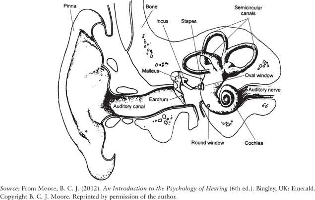
Lying on top of the basilar membrane are specialized sensory cells called hair cells. These run in rows along the length of the basilar membrane. One type of hair cell, called the inner hair cell, responds to the movement of the basilar membrane by generating an electrical signal that in turn leads to the release of a neurotransmitter that triggers activity in the neurons of the auditory nerve. Each neuron derives its response from the vibration at a specific place on the basilar membrane.
Information about the characteristics of sounds is carried in the auditory nerve in three basic ways:
- By the rate of firing of individual neurons, which will be referred to as the “amount” of neural activity. The more vibration there is at a given place, the greater is the amount of activity in neurons connected to that place. It is commonly believed that the subjective loudness of a sound is related to the amount of neural activity evoked by that sound, although this idea has been disputed.
- By the distribution of activity across neurons. Each neuron is tuned so that it responds most strongly to a specific frequency, called the characteristic frequency (CF); the tuning reflects the tuning of the place on the basilar membrane that drives the neuron. The distribution of the amount of neural activity as a function of CF is called the excitation pattern. The excitation pattern conveys “place” information since the CF at the peak of the excitation pattern is related to the place on the basilar membrane that is excited most.
- By the detailed time pattern of the neural impulses and especially the time intervals between successive nerve impulses. This form of information is known as “temporal” information. Neural impulses tend to be evoked at times corresponding to a specific phase of the waveform on the basilar membrane (for example, at the peaks of the waveform), an effect called phase locking. As a result, for a periodic sound, the time intervals between successive nerve impulses are approximately integer multiples of the period of the sound. For example, if the sound has a frequency of 500 hertz (Hz), the period is 2 milliseconds (ms), and the time intervals between successive nerve impulses would be close to 2, 4, 6, 8, 10, … ms. Phase locking breaks down at high frequencies (above about 36 kHz [kilohertz] in most mammals), but the upper limit in humans is not definitely known. Studies of pitch perception suggest that phase locking is very weak for frequencies above about 5 kHz.
In addition, information about sounds is conveyed by the differences between the two ears in all the above. In particular, differences in intensity at the two ears (primarily conveyed by differences in neural firing rate) and differences in the time of arrival of sounds at the two ears (conveyed mainly by subtle differences in the exact timing of nerve spikes) play a strong role in determining the perceived location of sounds.
...
- Action and Motor Control
- Action and Bodily Movement
- Analogical Reasoning, Models of Development
- Anomalous Monism
- Anomalous Monism
- Anti-Individualism About Cognition
- Anti-Individualism About Cognition
- Attention and Emotions, Computational Perspectives
- Belief and Judgment
- Case-Based Reasoning, Computational Perspectives
- Causal Theories of Intentionality
- Causal Theories of Memory
- Computational Models of Emotion
- Computational Perspectives
- Conscious Thinking
- Conscious Thinking
- Consciousness and Embodiment
- Consciousness and Embodiment
- Consciousness and the Unconscious
- Deductive Reasoning
- Descriptive Thought
- Development
- Disorders and Pathology
- Disorders and Pathology
- Distributed Cognition
- Electrophysiological Studies of Mind
- Eliminative Materialism
- Eliminative Materialism
- Emergence
- Emergence
- Emotion and Moral Judgment
- Evolutionary Perspectives
- Experimental Philosophy
- Explanatory Gap
- Explanatory Gap
- Extended Mind
- Face Recognition in Humans and Computers
- Facial Expressions, Computational Perspectives
- Facial Expressions, Computational Perspectives
- Freedom of Action
- Idealism
- Idealism
- Intelligence, Neural Basis
- Intentionality of Bodily Sensation
- Intentionality of Emotion
- Introspection
- Know-How, Philosophical Perspectives
- Know-How, Philosophical Perspectives
- Knowledge by Acquaintance
- Machine Speech Recognition
- Memory and Knowledge
- Mental Action
- Mental Causation
- Mental Causation
- Mind-Body Problem
- Modeling Causal Learning
- Moral Development
- Multimodal Conversational Systems
- Naïve Realism
- Natural Language Generation
- Neural Basis
- Object-Dependent Thought
- Personal Identity
- Personal Identity, Development of
- Philosophical Perspectives
- Philosophy of Action
- Physicalism
- Physicalism
- Psychological Research
- Realism and Instrumentalism
- Realism and Instrumentalism
- Reductive Physicalism
- Reductive Physicalism
- Relationships, Development of
- Representational Theory of Mind
- Self-Knowledge
- Semantic Memory, Computational Perspectives
- Sequential Memory, Computational Perspectives
- Serial Order Memory, Computational Perspectives
- Smell, Philosophical Perspectives
- Taste, Philosophical Perspectives
- Teleology
- Teleology
- Theory of Appearing
- Attention
- Aging, Memory, and Information Processing Speed
- Audition, Neural Basis
- Bilingual Language Processing
- Bilingualism, Cognitive Benefits of
- Black English Vernacular (Ebonics)
- Blindsight
- Borderline Personality Disorder
- Capgras Delusion
- Character and Personality, Philosophical Perspectives
- Collective Action
- Common Coding
- Computational Perspectives
- Distributed Cognition
- Emotion and Moral Judgment
- Emotion, Cultural Perspectives
- Emotional Recognition, Neuropsychology of
- Event Memory, Development
- Evolutionary Perspectives
- Experimental Philosophy
- Flynn Effect: Rising Intelligence Scores
- Gender Differences in Language and Language Use
- Gesture and Language Processing
- Attention and Action
- Attention and Emotion
- Attention, Resource Models
- Attentional Blink Effect
- Automaticity
- Change Blindness
- Divided Attention and Memory
- Inattentional Blindness
- Inhibition of Return
- Mental Effort
- Multitasking and Human Performance
- Neurodynamics of Visual Search
- Perceptual Consciousness and Attention
- Psychological Refractory Period
- Stroop Effect
- Visual Search
- Heritability
- Heritage Language and Second Language Learning
- Intelligence and Working Memory
- Joint or Collective Intention
- Joint or Collective Intention
- Knowledge Acquisition in Development
- Mirror Neurons
- Attention and Action
- Attention and Emotion
- Attention, Resource Models
- Attentional Blink Effect
- Automaticity
- Change Blindness
- Divided Attention and Memory
- Inattentional Blindness
- Inhibition of Return
- Mental Effort
- Multitasking and Human Performance
- Neurodynamics of Visual Search
- Perceptual Consciousness and Attention
- Psychological Refractory Period
- Stroop Effect
- Visual Search
- Multiple Intelligences Theory
- Narcissistic Personality Disorder
- Neural Basis
- Philosophical Perspectives
- Philosophy of Action
- Psychological Research
- Attention and Action
- Attention and Emotion
- Attention, Resource Models
- Attentional Blink Effect
- Automaticity
- Change Blindness
- Divided Attention and Memory
- Inattentional Blindness
- Inhibition of Return
- Mental Effort
- Multitasking and Human Performance
- Neurodynamics of Visual Search
- Perceptual Consciousness and Attention
- Psychological Refractory Period
- Stroop Effect
- Visual Search
- Self-Knowledge
- Synesthesia
- Visual Imagery
- Word Learning
- Concepts and Categories
- Amnesia
- Analogical Reasoning, Models of Development
- Anxiety Disorders
- Character and Personality, Philosophical Perspectives
- Computational Perspectives
- Confabulation
- Desire
- Development
- Disjunctive Theory of Perception
- Disorders and Pathology
- Dissent, Effects on Group Decisions
- Distributed Cognition
- Dyslexia, Developmental
- Dyslexia, Phonological Processing in
- Emotion and Moral Judgment
- Evolutionary Perspectives
- Exercise and the Brain
- Folk Psychology
- Group Decision Making
- Guilt
- Hearing, Philosophical Perspectives
- Heritage Language and Second Language Learning
- Human Classification Learning
- Innateness and Parameter Setting
- Intergroup Conflict
- Intergroup Conflict, Models of
- Knowledge Acquisition in Development
- Language Development
- Language Development, Overregulation in
- Moral Development
- Naïve Realism
- Neural Basis
- Philosophical Perspectives
- Political Psychology
- Prediction, Clinical Versus Actuarial
- Psychological Research
- Religion and Psychiatry
- Semantic Dementia
- Smell, Philosophical Perspectives
- Spatial Cognition, Development of
- Taste, Philosophical Perspectives
- Word Learning
- Consciousness
- Aphasia
- Attitude Change
- Attitude Change
- Attitudes and Behavior
- Attraction
- Attribution Theory
- Auditory Masking
- Behavioral Therapy
- Borderline Personality Disorder
- Cognitive Dissonance
- Anesthesia and Awareness
- Attention and Consciousness
- Consciousness and the Unconscious
- Metacognition and Education
- Perceptual Consciousness and Attention
- Self-Consciousness
- Self-Knowledge
- Subliminal Perception
- Unconscious Emotions, Psychological Perspectives
- Unconscious Perception
- Voluntary Action, Illusion of
- Common Coding
- Conduction Aphasia
- Consciousness, Comparative Perspectives
- Deductive Reasoning
- Depth Perception
- Desirable Difficulties Perspective on Learning
- Discrimination Learning, Training Methods
- Disorders and Pathology
- Dyslexia, Acquired
- Dyslexia, Developmental
- Dyslexia, Phonological Processing in
- Anesthesia and Awareness
- Attention and Consciousness
- Consciousness and the Unconscious
- Metacognition and Education
- Perceptual Consciousness and Attention
- Self-Consciousness
- Self-Knowledge
- Subliminal Perception
- Unconscious Emotions, Psychological Perspectives
- Unconscious Perception
- Voluntary Action, Illusion of
- Evolutionary Perspectives
- Exercise and the Brain
- Face Perception
- Facial Expressions, Emotional
- Anesthesia and Awareness
- Attention and Consciousness
- Consciousness and the Unconscious
- Metacognition and Education
- Perceptual Consciousness and Attention
- Self-Consciousness
- Self-Knowledge
- Subliminal Perception
- Unconscious Emotions, Psychological Perspectives
- Unconscious Perception
- Voluntary Action, Illusion of
- Group Decision Making
- Hearing
- Heritability
- Intergroup Conflict
- Intergroup Conflict, Models of
- Learning Styles
- Anesthesia and Awareness
- Attention and Consciousness
- Consciousness and the Unconscious
- Metacognition and Education
- Perceptual Consciousness and Attention
- Self-Consciousness
- Self-Knowledge
- Subliminal Perception
- Unconscious Emotions, Psychological Perspectives
- Unconscious Perception
- Voluntary Action, Illusion of
- Love
- McCollough Effect
- Meta-Analysis
- Metacognition and Education
- Motor Learning, Practical Aspects
- Multinomial Modeling
- Music Perception
- Neural Basis
- Optic Flow
- Perceptual Consciousness and Attention
- Perceptual Constancy
- Personal Identity, Development of
- Personality: Individual Versus Situation Debate
- Personality: Individual Versus Situation Debate
- Persuasion
- Philosophical Perspectives
- Placebo Effect
- Political Psychology
- Anesthesia and Awareness
- Attention and Consciousness
- Consciousness and the Unconscious
- Metacognition and Education
- Perceptual Consciousness and Attention
- Self-Consciousness
- Self-Knowledge
- Subliminal Perception
- Unconscious Emotions, Psychological Perspectives
- Unconscious Perception
- Voluntary Action, Illusion of
- Political Psychology
- Psychological Research
- Anesthesia and Awareness
- Attention and Consciousness
- Consciousness and the Unconscious
- Metacognition and Education
- Perceptual Consciousness and Attention
- Self-Consciousness
- Self-Knowledge
- Subliminal Perception
- Unconscious Emotions, Psychological Perspectives
- Unconscious Perception
- Voluntary Action, Illusion of
- Reaction Time
- Retrieval Practice (Testing) Effect
- Semantic Dementia
- Skill Learning, Enhancement of
- Social Cognition
- Social Loafing
- Spacing Effect, Practical Applications
- Speech Perception
- Stereopsis
- Subliminal Perception
- Synesthesia
- Time Perception
- Unconscious Perception
- Visual Imagery
- Visual Masking
- Visual Search
- Visuospatial Reasoning
- Wisdom of Crowds Effect
- Word Recognition, Auditory
- Word Recognition, Visual
- Working Memory, Evolution of
- Decision Making
- Amnesia
- Argument Mapping
- Behavioral Therapy
- Behaviorism
- Case-Based Reasoning, Computational Perspectives
- Computational Perspectives
- Desirable Difficulties Perspective on Learning
- Desire
- Desire
- Emotion and Psychopathology
- Emotion, Cerebral Lateralization
- Emotion, Psychophysiology of
- Emotional Recognition, Neuropsychology of
- Event Memory, Development
- Genes and Linguistic Tone
- Human Classification Learning
- Implicit Memory
- Learning Styles
- Memory, Neural Basis
- Metacognition and Education
- Music and the Evolution of Language
- Neural Basis
- Philosophical Perspectives
- Placebo Effect
- Practical Applications
- Psychological Research
- Reinforcement Learning, Psychological Perspectives
- Retrieval Practice (Testing) Effect
- Skill Learning, Enhancement of
- Spacing Effect
- Word Learning
- Disorders and Pathology
- Addiction
- Amnesia
- Analogical Mapping and Reasoning
- Anchoring
- Anosognosia
- Anxiety Disorders
- Aphasia
- Aphasia
- Apraxia
- Autism
- Automatic Behavior
- Autoscopic Phenomena
- Availability Heuristic
- Behavioral Therapy
- Borderline Personality Disorder
- Capgras Delusion
- Causal Theories of Memory
- Conduction Aphasia
- Conduction Aphasia
- Confabulation
- Delusions
- Desire
- Distributed Cognition
- Dyslexia, Acquired
- Dyslexia, Developmental
- Dyslexia, Phonological Processing in
- Emotion and Moral Judgment
- Emotion and Psychopathology
- Emotions and Consciousness
- Fregoli Delusion
- Hypochondria
- Intentionality of Emotion
- Legal Reasoning, Psychological Perspectives
- Memory and Knowledge
- Motivated Thinking
- Narcissistic Personality Disorder
- Objects of Memory
- Rationality of Emotion
- Religion and Psychiatry
- Representativeness Heuristic
- Schizophrenia
- Scientific Reasoning
- Semantic Dementia
- Spatial Cognition, Development of
- Thinking
- Two System Models of Reasoning
- Visual Imagery
- Visuospatial Reasoning
- Williams Syndrome
- Computational Perspectives
- Action and Bodily Movement
- Analogical Reasoning, Models of Development
- Anomalous Monism
- Anomalous Monism
- Anti-Individualism About Cognition
- Anti-Individualism About Cognition
- Attention and Emotions, Computational Perspectives
- Belief and Judgment
- Case-Based Reasoning, Computational Perspectives
- Causal Theories of Intentionality
- Causal Theories of Memory
- Computational Models of Emotion
- Computational Perspectives
- Conscious Thinking
- Conscious Thinking
- Consciousness and Embodiment
- Consciousness and Embodiment
- Consciousness and the Unconscious
- Deductive Reasoning
- Descriptive Thought
- Development
- Disorders and Pathology
- Disorders and Pathology
- Distributed Cognition
- Electrophysiological Studies of Mind
- Eliminative Materialism
- Eliminative Materialism
- Emergence
- Emergence
- Emotion and Moral Judgment
- Evolutionary Perspectives
- Experimental Philosophy
- Explanatory Gap
- Explanatory Gap
- Extended Mind
- Face Recognition in Humans and Computers
- Facial Expressions, Computational Perspectives
- Facial Expressions, Computational Perspectives
- Freedom of Action
- Idealism
- Idealism
- Intelligence, Neural Basis
- Intentionality of Bodily Sensation
- Intentionality of Emotion
- Introspection
- Know-How, Philosophical Perspectives
- Know-How, Philosophical Perspectives
- Knowledge by Acquaintance
- Machine Speech Recognition
- Memory and Knowledge
- Mental Action
- Mental Causation
- Mental Causation
- Mind-Body Problem
- Modeling Causal Learning
- Moral Development
- Multimodal Conversational Systems
- Naïve Realism
- Natural Language Generation
- Neural Basis
- Object-Dependent Thought
- Personal Identity
- Personal Identity, Development of
- Philosophical Perspectives
- Philosophy of Action
- Physicalism
- Physicalism
- Psychological Research
- Realism and Instrumentalism
- Realism and Instrumentalism
- Reductive Physicalism
- Reductive Physicalism
- Relationships, Development of
- Representational Theory of Mind
- Self-Knowledge
- Semantic Memory, Computational Perspectives
- Sequential Memory, Computational Perspectives
- Serial Order Memory, Computational Perspectives
- Smell, Philosophical Perspectives
- Taste, Philosophical Perspectives
- Teleology
- Teleology
- Theory of Appearing
- Cultural Perspectives
- Aging, Memory, and Information Processing Speed
- Audition, Neural Basis
- Bilingual Language Processing
- Bilingualism, Cognitive Benefits of
- Black English Vernacular (Ebonics)
- Blindsight
- Borderline Personality Disorder
- Capgras Delusion
- Character and Personality, Philosophical Perspectives
- Collective Action
- Common Coding
- Computational Perspectives
- Distributed Cognition
- Emotion and Moral Judgment
- Emotion, Cultural Perspectives
- Emotional Recognition, Neuropsychology of
- Event Memory, Development
- Evolutionary Perspectives
- Experimental Philosophy
- Flynn Effect: Rising Intelligence Scores
- Gender Differences in Language and Language Use
- Gesture and Language Processing
- Attention and Action
- Attention and Emotion
- Attention, Resource Models
- Attentional Blink Effect
- Automaticity
- Change Blindness
- Divided Attention and Memory
- Inattentional Blindness
- Inhibition of Return
- Mental Effort
- Multitasking and Human Performance
- Neurodynamics of Visual Search
- Perceptual Consciousness and Attention
- Psychological Refractory Period
- Stroop Effect
- Visual Search
- Heritability
- Heritage Language and Second Language Learning
- Intelligence and Working Memory
- Joint or Collective Intention
- Joint or Collective Intention
- Knowledge Acquisition in Development
- Mirror Neurons
- Attention and Action
- Attention and Emotion
- Attention, Resource Models
- Attentional Blink Effect
- Automaticity
- Change Blindness
- Divided Attention and Memory
- Inattentional Blindness
- Inhibition of Return
- Mental Effort
- Multitasking and Human Performance
- Neurodynamics of Visual Search
- Perceptual Consciousness and Attention
- Psychological Refractory Period
- Stroop Effect
- Visual Search
- Multiple Intelligences Theory
- Narcissistic Personality Disorder
- Neural Basis
- Philosophical Perspectives
- Philosophy of Action
- Psychological Research
- Attention and Action
- Attention and Emotion
- Attention, Resource Models
- Attentional Blink Effect
- Automaticity
- Change Blindness
- Divided Attention and Memory
- Inattentional Blindness
- Inhibition of Return
- Mental Effort
- Multitasking and Human Performance
- Neurodynamics of Visual Search
- Perceptual Consciousness and Attention
- Psychological Refractory Period
- Stroop Effect
- Visual Search
- Self-Knowledge
- Synesthesia
- Visual Imagery
- Word Learning
- Disorders and Pathology
- Amnesia
- Analogical Reasoning, Models of Development
- Anxiety Disorders
- Character and Personality, Philosophical Perspectives
- Computational Perspectives
- Confabulation
- Desire
- Development
- Disjunctive Theory of Perception
- Disorders and Pathology
- Dissent, Effects on Group Decisions
- Distributed Cognition
- Dyslexia, Developmental
- Dyslexia, Phonological Processing in
- Emotion and Moral Judgment
- Evolutionary Perspectives
- Exercise and the Brain
- Folk Psychology
- Group Decision Making
- Guilt
- Hearing, Philosophical Perspectives
- Heritage Language and Second Language Learning
- Human Classification Learning
- Innateness and Parameter Setting
- Intergroup Conflict
- Intergroup Conflict, Models of
- Knowledge Acquisition in Development
- Language Development
- Language Development, Overregulation in
- Moral Development
- Naïve Realism
- Neural Basis
- Philosophical Perspectives
- Political Psychology
- Prediction, Clinical Versus Actuarial
- Psychological Research
- Religion and Psychiatry
- Semantic Dementia
- Smell, Philosophical Perspectives
- Spatial Cognition, Development of
- Taste, Philosophical Perspectives
- Word Learning
- Evolutionary Perspectives
- Aphasia
- Attitude Change
- Attitude Change
- Attitudes and Behavior
- Attraction
- Attribution Theory
- Auditory Masking
- Behavioral Therapy
- Borderline Personality Disorder
- Cognitive Dissonance
- Anesthesia and Awareness
- Attention and Consciousness
- Consciousness and the Unconscious
- Metacognition and Education
- Perceptual Consciousness and Attention
- Self-Consciousness
- Self-Knowledge
- Subliminal Perception
- Unconscious Emotions, Psychological Perspectives
- Unconscious Perception
- Voluntary Action, Illusion of
- Common Coding
- Conduction Aphasia
- Consciousness, Comparative Perspectives
- Deductive Reasoning
- Depth Perception
- Desirable Difficulties Perspective on Learning
- Discrimination Learning, Training Methods
- Disorders and Pathology
- Dyslexia, Acquired
- Dyslexia, Developmental
- Dyslexia, Phonological Processing in
- Anesthesia and Awareness
- Attention and Consciousness
- Consciousness and the Unconscious
- Metacognition and Education
- Perceptual Consciousness and Attention
- Self-Consciousness
- Self-Knowledge
- Subliminal Perception
- Unconscious Emotions, Psychological Perspectives
- Unconscious Perception
- Voluntary Action, Illusion of
- Evolutionary Perspectives
- Exercise and the Brain
- Face Perception
- Facial Expressions, Emotional
- Anesthesia and Awareness
- Attention and Consciousness
- Consciousness and the Unconscious
- Metacognition and Education
- Perceptual Consciousness and Attention
- Self-Consciousness
- Self-Knowledge
- Subliminal Perception
- Unconscious Emotions, Psychological Perspectives
- Unconscious Perception
- Voluntary Action, Illusion of
- Group Decision Making
- Hearing
- Heritability
- Intergroup Conflict
- Intergroup Conflict, Models of
- Learning Styles
- Anesthesia and Awareness
- Attention and Consciousness
- Consciousness and the Unconscious
- Metacognition and Education
- Perceptual Consciousness and Attention
- Self-Consciousness
- Self-Knowledge
- Subliminal Perception
- Unconscious Emotions, Psychological Perspectives
- Unconscious Perception
- Voluntary Action, Illusion of
- Love
- McCollough Effect
- Meta-Analysis
- Metacognition and Education
- Motor Learning, Practical Aspects
- Multinomial Modeling
- Music Perception
- Neural Basis
- Optic Flow
- Perceptual Consciousness and Attention
- Perceptual Constancy
- Personal Identity, Development of
- Personality: Individual Versus Situation Debate
- Personality: Individual Versus Situation Debate
- Persuasion
- Philosophical Perspectives
- Placebo Effect
- Political Psychology
- Anesthesia and Awareness
- Attention and Consciousness
- Consciousness and the Unconscious
- Metacognition and Education
- Perceptual Consciousness and Attention
- Self-Consciousness
- Self-Knowledge
- Subliminal Perception
- Unconscious Emotions, Psychological Perspectives
- Unconscious Perception
- Voluntary Action, Illusion of
- Political Psychology
- Psychological Research
- Anesthesia and Awareness
- Attention and Consciousness
- Consciousness and the Unconscious
- Metacognition and Education
- Perceptual Consciousness and Attention
- Self-Consciousness
- Self-Knowledge
- Subliminal Perception
- Unconscious Emotions, Psychological Perspectives
- Unconscious Perception
- Voluntary Action, Illusion of
- Reaction Time
- Retrieval Practice (Testing) Effect
- Semantic Dementia
- Skill Learning, Enhancement of
- Social Cognition
- Social Loafing
- Spacing Effect, Practical Applications
- Speech Perception
- Stereopsis
- Subliminal Perception
- Synesthesia
- Time Perception
- Unconscious Perception
- Visual Imagery
- Visual Masking
- Visual Search
- Visuospatial Reasoning
- Wisdom of Crowds Effect
- Word Recognition, Auditory
- Word Recognition, Visual
- Working Memory, Evolution of
- Neural Basis
- Amnesia
- Argument Mapping
- Behavioral Therapy
- Behaviorism
- Case-Based Reasoning, Computational Perspectives
- Computational Perspectives
- Desirable Difficulties Perspective on Learning
- Desire
- Desire
- Emotion and Psychopathology
- Emotion, Cerebral Lateralization
- Emotion, Psychophysiology of
- Emotional Recognition, Neuropsychology of
- Event Memory, Development
- Genes and Linguistic Tone
- Human Classification Learning
- Implicit Memory
- Learning Styles
- Memory, Neural Basis
- Metacognition and Education
- Music and the Evolution of Language
- Neural Basis
- Philosophical Perspectives
- Placebo Effect
- Practical Applications
- Psychological Research
- Reinforcement Learning, Psychological Perspectives
- Retrieval Practice (Testing) Effect
- Skill Learning, Enhancement of
- Spacing Effect
- Word Learning
- Philosophical Perspectives
- Addiction
- Amnesia
- Analogical Mapping and Reasoning
- Anchoring
- Anosognosia
- Anxiety Disorders
- Aphasia
- Aphasia
- Apraxia
- Autism
- Automatic Behavior
- Autoscopic Phenomena
- Availability Heuristic
- Behavioral Therapy
- Borderline Personality Disorder
- Capgras Delusion
- Causal Theories of Memory
- Conduction Aphasia
- Conduction Aphasia
- Confabulation
- Delusions
- Desire
- Distributed Cognition
- Dyslexia, Acquired
- Dyslexia, Developmental
- Dyslexia, Phonological Processing in
- Emotion and Moral Judgment
- Emotion and Psychopathology
- Emotions and Consciousness
- Fregoli Delusion
- Hypochondria
- Intentionality of Emotion
- Legal Reasoning, Psychological Perspectives
- Memory and Knowledge
- Motivated Thinking
- Narcissistic Personality Disorder
- Objects of Memory
- Rationality of Emotion
- Religion and Psychiatry
- Representativeness Heuristic
- Schizophrenia
- Scientific Reasoning
- Semantic Dementia
- Spatial Cognition, Development of
- Thinking
- Two System Models of Reasoning
- Visual Imagery
- Visuospatial Reasoning
- Williams Syndrome
- Psychological Research
- Affective Forecasting
- Anxiety Disorders
- Attention and Emotion
- Cognitive Dissonance
- Descriptions
- Descriptive Thought
- Disgust
- Embarrassment
- Emotion and Moral Judgment
- Emotion and Working Memory
- Emotion Regulation
- Emotion, Psychophysiology of
- Emotion, Structural Approaches
- Envy
- Exercise and the Brain
- Facial Expressions, Emotional
- Guilt
- Happiness
- Indexical Thought
- Intension and Extension
- Jealousy
- Love
- Metaphor
- Mnemonic Strategies
- Object-Dependent Thought
- Regret
- Religion and Psychiatry
- Resentment
- Retrieval Practice (Testing) Effect
- Spacing Effect, Practical Applications
- Unconscious Emotions, Psychological Perspectives
- Philosophical Perspectives
- Action and Bodily Movement
- Analogical Reasoning, Models of Development
- Anomalous Monism
- Anomalous Monism
- Anti-Individualism About Cognition
- Anti-Individualism About Cognition
- Attention and Emotions, Computational Perspectives
- Belief and Judgment
- Case-Based Reasoning, Computational Perspectives
- Causal Theories of Intentionality
- Causal Theories of Memory
- Computational Models of Emotion
- Computational Perspectives
- Conscious Thinking
- Conscious Thinking
- Consciousness and Embodiment
- Consciousness and Embodiment
- Consciousness and the Unconscious
- Deductive Reasoning
- Descriptive Thought
- Development
- Disorders and Pathology
- Disorders and Pathology
- Distributed Cognition
- Electrophysiological Studies of Mind
- Eliminative Materialism
- Eliminative Materialism
- Emergence
- Emergence
- Emotion and Moral Judgment
- Evolutionary Perspectives
- Experimental Philosophy
- Explanatory Gap
- Explanatory Gap
- Extended Mind
- Face Recognition in Humans and Computers
- Facial Expressions, Computational Perspectives
- Facial Expressions, Computational Perspectives
- Freedom of Action
- Idealism
- Idealism
- Intelligence, Neural Basis
- Intentionality of Bodily Sensation
- Intentionality of Emotion
- Introspection
- Know-How, Philosophical Perspectives
- Know-How, Philosophical Perspectives
- Knowledge by Acquaintance
- Machine Speech Recognition
- Memory and Knowledge
- Mental Action
- Mental Causation
- Mental Causation
- Mind-Body Problem
- Modeling Causal Learning
- Moral Development
- Multimodal Conversational Systems
- Naïve Realism
- Natural Language Generation
- Neural Basis
- Object-Dependent Thought
- Personal Identity
- Personal Identity, Development of
- Philosophical Perspectives
- Philosophy of Action
- Physicalism
- Physicalism
- Psychological Research
- Realism and Instrumentalism
- Realism and Instrumentalism
- Reductive Physicalism
- Reductive Physicalism
- Relationships, Development of
- Representational Theory of Mind
- Self-Knowledge
- Semantic Memory, Computational Perspectives
- Sequential Memory, Computational Perspectives
- Serial Order Memory, Computational Perspectives
- Smell, Philosophical Perspectives
- Taste, Philosophical Perspectives
- Teleology
- Teleology
- Theory of Appearing
- Psychological Research
- Aging, Memory, and Information Processing Speed
- Audition, Neural Basis
- Bilingual Language Processing
- Bilingualism, Cognitive Benefits of
- Black English Vernacular (Ebonics)
- Blindsight
- Borderline Personality Disorder
- Capgras Delusion
- Character and Personality, Philosophical Perspectives
- Collective Action
- Common Coding
- Computational Perspectives
- Distributed Cognition
- Emotion and Moral Judgment
- Emotion, Cultural Perspectives
- Emotional Recognition, Neuropsychology of
- Event Memory, Development
- Evolutionary Perspectives
- Experimental Philosophy
- Flynn Effect: Rising Intelligence Scores
- Gender Differences in Language and Language Use
- Gesture and Language Processing
- Attention and Action
- Attention and Emotion
- Attention, Resource Models
- Attentional Blink Effect
- Automaticity
- Change Blindness
- Divided Attention and Memory
- Inattentional Blindness
- Inhibition of Return
- Mental Effort
- Multitasking and Human Performance
- Neurodynamics of Visual Search
- Perceptual Consciousness and Attention
- Psychological Refractory Period
- Stroop Effect
- Visual Search
- Heritability
- Heritage Language and Second Language Learning
- Intelligence and Working Memory
- Joint or Collective Intention
- Joint or Collective Intention
- Knowledge Acquisition in Development
- Mirror Neurons
- Attention and Action
- Attention and Emotion
- Attention, Resource Models
- Attentional Blink Effect
- Automaticity
- Change Blindness
- Divided Attention and Memory
- Inattentional Blindness
- Inhibition of Return
- Mental Effort
- Multitasking and Human Performance
- Neurodynamics of Visual Search
- Perceptual Consciousness and Attention
- Psychological Refractory Period
- Stroop Effect
- Visual Search
- Multiple Intelligences Theory
- Narcissistic Personality Disorder
- Neural Basis
- Philosophical Perspectives
- Philosophy of Action
- Psychological Research
- Attention and Action
- Attention and Emotion
- Attention, Resource Models
- Attentional Blink Effect
- Automaticity
- Change Blindness
- Divided Attention and Memory
- Inattentional Blindness
- Inhibition of Return
- Mental Effort
- Multitasking and Human Performance
- Neurodynamics of Visual Search
- Perceptual Consciousness and Attention
- Psychological Refractory Period
- Stroop Effect
- Visual Search
- Self-Knowledge
- Synesthesia
- Visual Imagery
- Word Learning
- Heritability
- Action and Bodily Movement
- Analogical Reasoning, Models of Development
- Anomalous Monism
- Anomalous Monism
- Anti-Individualism About Cognition
- Anti-Individualism About Cognition
- Attention and Emotions, Computational Perspectives
- Belief and Judgment
- Case-Based Reasoning, Computational Perspectives
- Causal Theories of Intentionality
- Causal Theories of Memory
- Computational Models of Emotion
- Computational Perspectives
- Conscious Thinking
- Conscious Thinking
- Consciousness and Embodiment
- Consciousness and Embodiment
- Consciousness and the Unconscious
- Deductive Reasoning
- Descriptive Thought
- Development
- Disorders and Pathology
- Disorders and Pathology
- Distributed Cognition
- Electrophysiological Studies of Mind
- Eliminative Materialism
- Eliminative Materialism
- Emergence
- Emergence
- Emotion and Moral Judgment
- Evolutionary Perspectives
- Experimental Philosophy
- Explanatory Gap
- Explanatory Gap
- Extended Mind
- Face Recognition in Humans and Computers
- Facial Expressions, Computational Perspectives
- Facial Expressions, Computational Perspectives
- Freedom of Action
- Idealism
- Idealism
- Intelligence, Neural Basis
- Intentionality of Bodily Sensation
- Intentionality of Emotion
- Introspection
- Know-How, Philosophical Perspectives
- Know-How, Philosophical Perspectives
- Knowledge by Acquaintance
- Machine Speech Recognition
- Memory and Knowledge
- Mental Action
- Mental Causation
- Mental Causation
- Mind-Body Problem
- Modeling Causal Learning
- Moral Development
- Multimodal Conversational Systems
- Naïve Realism
- Natural Language Generation
- Neural Basis
- Object-Dependent Thought
- Personal Identity
- Personal Identity, Development of
- Philosophical Perspectives
- Philosophy of Action
- Physicalism
- Physicalism
- Psychological Research
- Realism and Instrumentalism
- Realism and Instrumentalism
- Reductive Physicalism
- Reductive Physicalism
- Relationships, Development of
- Representational Theory of Mind
- Self-Knowledge
- Semantic Memory, Computational Perspectives
- Sequential Memory, Computational Perspectives
- Serial Order Memory, Computational Perspectives
- Smell, Philosophical Perspectives
- Taste, Philosophical Perspectives
- Teleology
- Teleology
- Theory of Appearing
- Cultural Perspectives
- Action and Bodily Movement
- Analogical Reasoning, Models of Development
- Anomalous Monism
- Anomalous Monism
- Anti-Individualism About Cognition
- Anti-Individualism About Cognition
- Attention and Emotions, Computational Perspectives
- Belief and Judgment
- Case-Based Reasoning, Computational Perspectives
- Causal Theories of Intentionality
- Causal Theories of Memory
- Computational Models of Emotion
- Computational Perspectives
- Conscious Thinking
- Conscious Thinking
- Consciousness and Embodiment
- Consciousness and Embodiment
- Consciousness and the Unconscious
- Deductive Reasoning
- Descriptive Thought
- Development
- Disorders and Pathology
- Disorders and Pathology
- Distributed Cognition
- Electrophysiological Studies of Mind
- Eliminative Materialism
- Eliminative Materialism
- Emergence
- Emergence
- Emotion and Moral Judgment
- Evolutionary Perspectives
- Experimental Philosophy
- Explanatory Gap
- Explanatory Gap
- Extended Mind
- Face Recognition in Humans and Computers
- Facial Expressions, Computational Perspectives
- Facial Expressions, Computational Perspectives
- Freedom of Action
- Idealism
- Idealism
- Intelligence, Neural Basis
- Intentionality of Bodily Sensation
- Intentionality of Emotion
- Introspection
- Know-How, Philosophical Perspectives
- Know-How, Philosophical Perspectives
- Knowledge by Acquaintance
- Machine Speech Recognition
- Memory and Knowledge
- Mental Action
- Mental Causation
- Mental Causation
- Mind-Body Problem
- Modeling Causal Learning
- Moral Development
- Multimodal Conversational Systems
- Naïve Realism
- Natural Language Generation
- Neural Basis
- Object-Dependent Thought
- Personal Identity
- Personal Identity, Development of
- Philosophical Perspectives
- Philosophy of Action
- Physicalism
- Physicalism
- Psychological Research
- Realism and Instrumentalism
- Realism and Instrumentalism
- Reductive Physicalism
- Reductive Physicalism
- Relationships, Development of
- Representational Theory of Mind
- Self-Knowledge
- Semantic Memory, Computational Perspectives
- Sequential Memory, Computational Perspectives
- Serial Order Memory, Computational Perspectives
- Smell, Philosophical Perspectives
- Taste, Philosophical Perspectives
- Teleology
- Teleology
- Theory of Appearing
- Philosophical Perspectives
- Aging, Memory, and Information Processing Speed
- Audition, Neural Basis
- Bilingual Language Processing
- Bilingualism, Cognitive Benefits of
- Black English Vernacular (Ebonics)
- Blindsight
- Borderline Personality Disorder
- Capgras Delusion
- Character and Personality, Philosophical Perspectives
- Collective Action
- Common Coding
- Computational Perspectives
- Distributed Cognition
- Emotion and Moral Judgment
- Emotion, Cultural Perspectives
- Emotional Recognition, Neuropsychology of
- Event Memory, Development
- Evolutionary Perspectives
- Experimental Philosophy
- Flynn Effect: Rising Intelligence Scores
- Gender Differences in Language and Language Use
- Gesture and Language Processing
- Attention and Action
- Attention and Emotion
- Attention, Resource Models
- Attentional Blink Effect
- Automaticity
- Change Blindness
- Divided Attention and Memory
- Inattentional Blindness
- Inhibition of Return
- Mental Effort
- Multitasking and Human Performance
- Neurodynamics of Visual Search
- Perceptual Consciousness and Attention
- Psychological Refractory Period
- Stroop Effect
- Visual Search
- Heritability
- Heritage Language and Second Language Learning
- Intelligence and Working Memory
- Joint or Collective Intention
- Joint or Collective Intention
- Knowledge Acquisition in Development
- Mirror Neurons
- Attention and Action
- Attention and Emotion
- Attention, Resource Models
- Attentional Blink Effect
- Automaticity
- Change Blindness
- Divided Attention and Memory
- Inattentional Blindness
- Inhibition of Return
- Mental Effort
- Multitasking and Human Performance
- Neurodynamics of Visual Search
- Perceptual Consciousness and Attention
- Psychological Refractory Period
- Stroop Effect
- Visual Search
- Multiple Intelligences Theory
- Narcissistic Personality Disorder
- Neural Basis
- Philosophical Perspectives
- Philosophy of Action
- Psychological Research
- Attention and Action
- Attention and Emotion
- Attention, Resource Models
- Attentional Blink Effect
- Automaticity
- Change Blindness
- Divided Attention and Memory
- Inattentional Blindness
- Inhibition of Return
- Mental Effort
- Multitasking and Human Performance
- Neurodynamics of Visual Search
- Perceptual Consciousness and Attention
- Psychological Refractory Period
- Stroop Effect
- Visual Search
- Self-Knowledge
- Synesthesia
- Visual Imagery
- Word Learning
- Psychological Research
- Amnesia
- Analogical Reasoning, Models of Development
- Anxiety Disorders
- Character and Personality, Philosophical Perspectives
- Computational Perspectives
- Confabulation
- Desire
- Development
- Disjunctive Theory of Perception
- Disorders and Pathology
- Dissent, Effects on Group Decisions
- Distributed Cognition
- Dyslexia, Developmental
- Dyslexia, Phonological Processing in
- Emotion and Moral Judgment
- Evolutionary Perspectives
- Exercise and the Brain
- Folk Psychology
- Group Decision Making
- Guilt
- Hearing, Philosophical Perspectives
- Heritage Language and Second Language Learning
- Human Classification Learning
- Innateness and Parameter Setting
- Intergroup Conflict
- Intergroup Conflict, Models of
- Knowledge Acquisition in Development
- Language Development
- Language Development, Overregulation in
- Moral Development
- Naïve Realism
- Neural Basis
- Philosophical Perspectives
- Political Psychology
- Prediction, Clinical Versus Actuarial
- Psychological Research
- Religion and Psychiatry
- Semantic Dementia
- Smell, Philosophical Perspectives
- Spatial Cognition, Development of
- Taste, Philosophical Perspectives
- Word Learning
- Neural Basis
- Action and Bodily Movement
- Analogical Reasoning, Models of Development
- Anomalous Monism
- Anomalous Monism
- Anti-Individualism About Cognition
- Anti-Individualism About Cognition
- Attention and Emotions, Computational Perspectives
- Belief and Judgment
- Case-Based Reasoning, Computational Perspectives
- Causal Theories of Intentionality
- Causal Theories of Memory
- Computational Models of Emotion
- Computational Perspectives
- Conscious Thinking
- Conscious Thinking
- Consciousness and Embodiment
- Consciousness and Embodiment
- Consciousness and the Unconscious
- Deductive Reasoning
- Descriptive Thought
- Development
- Disorders and Pathology
- Disorders and Pathology
- Distributed Cognition
- Electrophysiological Studies of Mind
- Eliminative Materialism
- Eliminative Materialism
- Emergence
- Emergence
- Emotion and Moral Judgment
- Evolutionary Perspectives
- Experimental Philosophy
- Explanatory Gap
- Explanatory Gap
- Extended Mind
- Face Recognition in Humans and Computers
- Facial Expressions, Computational Perspectives
- Facial Expressions, Computational Perspectives
- Freedom of Action
- Idealism
- Idealism
- Intelligence, Neural Basis
- Intentionality of Bodily Sensation
- Intentionality of Emotion
- Introspection
- Know-How, Philosophical Perspectives
- Know-How, Philosophical Perspectives
- Knowledge by Acquaintance
- Machine Speech Recognition
- Memory and Knowledge
- Mental Action
- Mental Causation
- Mental Causation
- Mind-Body Problem
- Modeling Causal Learning
- Moral Development
- Multimodal Conversational Systems
- Naïve Realism
- Natural Language Generation
- Neural Basis
- Object-Dependent Thought
- Personal Identity
- Personal Identity, Development of
- Philosophical Perspectives
- Philosophy of Action
- Physicalism
- Physicalism
- Psychological Research
- Realism and Instrumentalism
- Realism and Instrumentalism
- Reductive Physicalism
- Reductive Physicalism
- Relationships, Development of
- Representational Theory of Mind
- Self-Knowledge
- Semantic Memory, Computational Perspectives
- Sequential Memory, Computational Perspectives
- Serial Order Memory, Computational Perspectives
- Smell, Philosophical Perspectives
- Taste, Philosophical Perspectives
- Teleology
- Teleology
- Theory of Appearing
- Psychological Research
- Aging, Memory, and Information Processing Speed
- Audition, Neural Basis
- Bilingual Language Processing
- Bilingualism, Cognitive Benefits of
- Black English Vernacular (Ebonics)
- Blindsight
- Borderline Personality Disorder
- Capgras Delusion
- Character and Personality, Philosophical Perspectives
- Collective Action
- Common Coding
- Computational Perspectives
- Distributed Cognition
- Emotion and Moral Judgment
- Emotion, Cultural Perspectives
- Emotional Recognition, Neuropsychology of
- Event Memory, Development
- Evolutionary Perspectives
- Experimental Philosophy
- Flynn Effect: Rising Intelligence Scores
- Gender Differences in Language and Language Use
- Gesture and Language Processing
- Attention and Action
- Attention and Emotion
- Attention, Resource Models
- Attentional Blink Effect
- Automaticity
- Change Blindness
- Divided Attention and Memory
- Inattentional Blindness
- Inhibition of Return
- Mental Effort
- Multitasking and Human Performance
- Neurodynamics of Visual Search
- Perceptual Consciousness and Attention
- Psychological Refractory Period
- Stroop Effect
- Visual Search
- Heritability
- Heritage Language and Second Language Learning
- Intelligence and Working Memory
- Joint or Collective Intention
- Joint or Collective Intention
- Knowledge Acquisition in Development
- Mirror Neurons
- Attention and Action
- Attention and Emotion
- Attention, Resource Models
- Attentional Blink Effect
- Automaticity
- Change Blindness
- Divided Attention and Memory
- Inattentional Blindness
- Inhibition of Return
- Mental Effort
- Multitasking and Human Performance
- Neurodynamics of Visual Search
- Perceptual Consciousness and Attention
- Psychological Refractory Period
- Stroop Effect
- Visual Search
- Multiple Intelligences Theory
- Narcissistic Personality Disorder
- Neural Basis
- Philosophical Perspectives
- Philosophy of Action
- Psychological Research
- Attention and Action
- Attention and Emotion
- Attention, Resource Models
- Attentional Blink Effect
- Automaticity
- Change Blindness
- Divided Attention and Memory
- Inattentional Blindness
- Inhibition of Return
- Mental Effort
- Multitasking and Human Performance
- Neurodynamics of Visual Search
- Perceptual Consciousness and Attention
- Psychological Refractory Period
- Stroop Effect
- Visual Search
- Self-Knowledge
- Synesthesia
- Visual Imagery
- Word Learning
- Computational Perspectives
- Action and Bodily Movement
- Analogical Reasoning, Models of Development
- Anomalous Monism
- Anomalous Monism
- Anti-Individualism About Cognition
- Anti-Individualism About Cognition
- Attention and Emotions, Computational Perspectives
- Belief and Judgment
- Case-Based Reasoning, Computational Perspectives
- Causal Theories of Intentionality
- Causal Theories of Memory
- Computational Models of Emotion
- Computational Perspectives
- Conscious Thinking
- Conscious Thinking
- Consciousness and Embodiment
- Consciousness and Embodiment
- Consciousness and the Unconscious
- Deductive Reasoning
- Descriptive Thought
- Development
- Disorders and Pathology
- Disorders and Pathology
- Distributed Cognition
- Electrophysiological Studies of Mind
- Eliminative Materialism
- Eliminative Materialism
- Emergence
- Emergence
- Emotion and Moral Judgment
- Evolutionary Perspectives
- Experimental Philosophy
- Explanatory Gap
- Explanatory Gap
- Extended Mind
- Face Recognition in Humans and Computers
- Facial Expressions, Computational Perspectives
- Facial Expressions, Computational Perspectives
- Freedom of Action
- Idealism
- Idealism
- Intelligence, Neural Basis
- Intentionality of Bodily Sensation
- Intentionality of Emotion
- Introspection
- Know-How, Philosophical Perspectives
- Know-How, Philosophical Perspectives
- Knowledge by Acquaintance
- Machine Speech Recognition
- Memory and Knowledge
- Mental Action
- Mental Causation
- Mental Causation
- Mind-Body Problem
- Modeling Causal Learning
- Moral Development
- Multimodal Conversational Systems
- Naïve Realism
- Natural Language Generation
- Neural Basis
- Object-Dependent Thought
- Personal Identity
- Personal Identity, Development of
- Philosophical Perspectives
- Philosophy of Action
- Physicalism
- Physicalism
- Psychological Research
- Realism and Instrumentalism
- Realism and Instrumentalism
- Reductive Physicalism
- Reductive Physicalism
- Relationships, Development of
- Representational Theory of Mind
- Self-Knowledge
- Semantic Memory, Computational Perspectives
- Sequential Memory, Computational Perspectives
- Serial Order Memory, Computational Perspectives
- Smell, Philosophical Perspectives
- Taste, Philosophical Perspectives
- Teleology
- Teleology
- Theory of Appearing
- Cultural Perspectives
- Aging, Memory, and Information Processing Speed
- Audition, Neural Basis
- Bilingual Language Processing
- Bilingualism, Cognitive Benefits of
- Black English Vernacular (Ebonics)
- Blindsight
- Borderline Personality Disorder
- Capgras Delusion
- Character and Personality, Philosophical Perspectives
- Collective Action
- Common Coding
- Computational Perspectives
- Distributed Cognition
- Emotion and Moral Judgment
- Emotion, Cultural Perspectives
- Emotional Recognition, Neuropsychology of
- Event Memory, Development
- Evolutionary Perspectives
- Experimental Philosophy
- Flynn Effect: Rising Intelligence Scores
- Gender Differences in Language and Language Use
- Gesture and Language Processing
- Attention and Action
- Attention and Emotion
- Attention, Resource Models
- Attentional Blink Effect
- Automaticity
- Change Blindness
- Divided Attention and Memory
- Inattentional Blindness
- Inhibition of Return
- Mental Effort
- Multitasking and Human Performance
- Neurodynamics of Visual Search
- Perceptual Consciousness and Attention
- Psychological Refractory Period
- Stroop Effect
- Visual Search
- Heritability
- Heritage Language and Second Language Learning
- Intelligence and Working Memory
- Joint or Collective Intention
- Joint or Collective Intention
- Knowledge Acquisition in Development
- Mirror Neurons
- Attention and Action
- Attention and Emotion
- Attention, Resource Models
- Attentional Blink Effect
- Automaticity
- Change Blindness
- Divided Attention and Memory
- Inattentional Blindness
- Inhibition of Return
- Mental Effort
- Multitasking and Human Performance
- Neurodynamics of Visual Search
- Perceptual Consciousness and Attention
- Psychological Refractory Period
- Stroop Effect
- Visual Search
- Multiple Intelligences Theory
- Narcissistic Personality Disorder
- Neural Basis
- Philosophical Perspectives
- Philosophy of Action
- Psychological Research
- Attention and Action
- Attention and Emotion
- Attention, Resource Models
- Attentional Blink Effect
- Automaticity
- Change Blindness
- Divided Attention and Memory
- Inattentional Blindness
- Inhibition of Return
- Mental Effort
- Multitasking and Human Performance
- Neurodynamics of Visual Search
- Perceptual Consciousness and Attention
- Psychological Refractory Period
- Stroop Effect
- Visual Search
- Self-Knowledge
- Synesthesia
- Visual Imagery
- Word Learning
- Development
- Amnesia
- Analogical Reasoning, Models of Development
- Anxiety Disorders
- Character and Personality, Philosophical Perspectives
- Computational Perspectives
- Confabulation
- Desire
- Development
- Disjunctive Theory of Perception
- Disorders and Pathology
- Dissent, Effects on Group Decisions
- Distributed Cognition
- Dyslexia, Developmental
- Dyslexia, Phonological Processing in
- Emotion and Moral Judgment
- Evolutionary Perspectives
- Exercise and the Brain
- Folk Psychology
- Group Decision Making
- Guilt
- Hearing, Philosophical Perspectives
- Heritage Language and Second Language Learning
- Human Classification Learning
- Innateness and Parameter Setting
- Intergroup Conflict
- Intergroup Conflict, Models of
- Knowledge Acquisition in Development
- Language Development
- Language Development, Overregulation in
- Moral Development
- Naïve Realism
- Neural Basis
- Philosophical Perspectives
- Political Psychology
- Prediction, Clinical Versus Actuarial
- Psychological Research
- Religion and Psychiatry
- Semantic Dementia
- Smell, Philosophical Perspectives
- Spatial Cognition, Development of
- Taste, Philosophical Perspectives
- Word Learning
- Disorders and Pathology
- Aphasia
- Attitude Change
- Attitude Change
- Attitudes and Behavior
- Attraction
- Attribution Theory
- Auditory Masking
- Behavioral Therapy
- Borderline Personality Disorder
- Cognitive Dissonance
- Anesthesia and Awareness
- Attention and Consciousness
- Consciousness and the Unconscious
- Metacognition and Education
- Perceptual Consciousness and Attention
- Self-Consciousness
- Self-Knowledge
- Subliminal Perception
- Unconscious Emotions, Psychological Perspectives
- Unconscious Perception
- Voluntary Action, Illusion of
- Common Coding
- Conduction Aphasia
- Consciousness, Comparative Perspectives
- Deductive Reasoning
- Depth Perception
- Desirable Difficulties Perspective on Learning
- Discrimination Learning, Training Methods
- Disorders and Pathology
- Dyslexia, Acquired
- Dyslexia, Developmental
- Dyslexia, Phonological Processing in
- Anesthesia and Awareness
- Attention and Consciousness
- Consciousness and the Unconscious
- Metacognition and Education
- Perceptual Consciousness and Attention
- Self-Consciousness
- Self-Knowledge
- Subliminal Perception
- Unconscious Emotions, Psychological Perspectives
- Unconscious Perception
- Voluntary Action, Illusion of
- Evolutionary Perspectives
- Exercise and the Brain
- Face Perception
- Facial Expressions, Emotional
- Anesthesia and Awareness
- Attention and Consciousness
- Consciousness and the Unconscious
- Metacognition and Education
- Perceptual Consciousness and Attention
- Self-Consciousness
- Self-Knowledge
- Subliminal Perception
- Unconscious Emotions, Psychological Perspectives
- Unconscious Perception
- Voluntary Action, Illusion of
- Group Decision Making
- Hearing
- Heritability
- Intergroup Conflict
- Intergroup Conflict, Models of
- Learning Styles
- Anesthesia and Awareness
- Attention and Consciousness
- Consciousness and the Unconscious
- Metacognition and Education
- Perceptual Consciousness and Attention
- Self-Consciousness
- Self-Knowledge
- Subliminal Perception
- Unconscious Emotions, Psychological Perspectives
- Unconscious Perception
- Voluntary Action, Illusion of
- Love
- McCollough Effect
- Meta-Analysis
- Metacognition and Education
- Motor Learning, Practical Aspects
- Multinomial Modeling
- Music Perception
- Neural Basis
- Optic Flow
- Perceptual Consciousness and Attention
- Perceptual Constancy
- Personal Identity, Development of
- Personality: Individual Versus Situation Debate
- Personality: Individual Versus Situation Debate
- Persuasion
- Philosophical Perspectives
- Placebo Effect
- Political Psychology
- Anesthesia and Awareness
- Attention and Consciousness
- Consciousness and the Unconscious
- Metacognition and Education
- Perceptual Consciousness and Attention
- Self-Consciousness
- Self-Knowledge
- Subliminal Perception
- Unconscious Emotions, Psychological Perspectives
- Unconscious Perception
- Voluntary Action, Illusion of
- Political Psychology
- Psychological Research
- Anesthesia and Awareness
- Attention and Consciousness
- Consciousness and the Unconscious
- Metacognition and Education
- Perceptual Consciousness and Attention
- Self-Consciousness
- Self-Knowledge
- Subliminal Perception
- Unconscious Emotions, Psychological Perspectives
- Unconscious Perception
- Voluntary Action, Illusion of
- Reaction Time
- Retrieval Practice (Testing) Effect
- Semantic Dementia
- Skill Learning, Enhancement of
- Social Cognition
- Social Loafing
- Spacing Effect, Practical Applications
- Speech Perception
- Stereopsis
- Subliminal Perception
- Synesthesia
- Time Perception
- Unconscious Perception
- Visual Imagery
- Visual Masking
- Visual Search
- Visuospatial Reasoning
- Wisdom of Crowds Effect
- Word Recognition, Auditory
- Word Recognition, Visual
- Working Memory, Evolution of
- Evolutionary Perspectives
- Amnesia
- Argument Mapping
- Behavioral Therapy
- Behaviorism
- Case-Based Reasoning, Computational Perspectives
- Computational Perspectives
- Desirable Difficulties Perspective on Learning
- Desire
- Desire
- Emotion and Psychopathology
- Emotion, Cerebral Lateralization
- Emotion, Psychophysiology of
- Emotional Recognition, Neuropsychology of
- Event Memory, Development
- Genes and Linguistic Tone
- Human Classification Learning
- Implicit Memory
- Learning Styles
- Memory, Neural Basis
- Metacognition and Education
- Music and the Evolution of Language
- Neural Basis
- Philosophical Perspectives
- Placebo Effect
- Practical Applications
- Psychological Research
- Reinforcement Learning, Psychological Perspectives
- Retrieval Practice (Testing) Effect
- Skill Learning, Enhancement of
- Spacing Effect
- Word Learning
- Neural Basis
- Addiction
- Amnesia
- Analogical Mapping and Reasoning
- Anchoring
- Anosognosia
- Anxiety Disorders
- Aphasia
- Aphasia
- Apraxia
- Autism
- Automatic Behavior
- Autoscopic Phenomena
- Availability Heuristic
- Behavioral Therapy
- Borderline Personality Disorder
- Capgras Delusion
- Causal Theories of Memory
- Conduction Aphasia
- Conduction Aphasia
- Confabulation
- Delusions
- Desire
- Distributed Cognition
- Dyslexia, Acquired
- Dyslexia, Developmental
- Dyslexia, Phonological Processing in
- Emotion and Moral Judgment
- Emotion and Psychopathology
- Emotions and Consciousness
- Fregoli Delusion
- Hypochondria
- Intentionality of Emotion
- Legal Reasoning, Psychological Perspectives
- Memory and Knowledge
- Motivated Thinking
- Narcissistic Personality Disorder
- Objects of Memory
- Rationality of Emotion
- Religion and Psychiatry
- Representativeness Heuristic
- Schizophrenia
- Scientific Reasoning
- Semantic Dementia
- Spatial Cognition, Development of
- Thinking
- Two System Models of Reasoning
- Visual Imagery
- Visuospatial Reasoning
- Williams Syndrome
- Philosophical Perspectives
- Affective Forecasting
- Anxiety Disorders
- Attention and Emotion
- Cognitive Dissonance
- Descriptions
- Descriptive Thought
- Disgust
- Embarrassment
- Emotion and Moral Judgment
- Emotion and Working Memory
- Emotion Regulation
- Emotion, Psychophysiology of
- Emotion, Structural Approaches
- Envy
- Exercise and the Brain
- Facial Expressions, Emotional
- Guilt
- Happiness
- Indexical Thought
- Intension and Extension
- Jealousy
- Love
- Metaphor
- Mnemonic Strategies
- Object-Dependent Thought
- Regret
- Religion and Psychiatry
- Resentment
- Retrieval Practice (Testing) Effect
- Spacing Effect, Practical Applications
- Unconscious Emotions, Psychological Perspectives
- Practical Applications
- Aging, Memory, and Information Processing Speed
- Deception, Linguistic Cues to
- Divided Attention and Memory
- Emotion and Working Memory
- Event Memory, Development
- Exercise and the Brain
- Eyewitness Memory
- Implicit Memory
- Intelligence and Working Memory
- Lie Detection
- Memory Recall, Dynamics
- Memory, Interference With
- Rehearsal and Memory
- Retrieval Practice (Testing) Effect
- Semantic Memory
- Spacing Effect
- Visual Working Memory
- Working Memory
- Working Memory in Language Processing
- Psychological Research
- Bilingual Language Processing
- Bilingualism, Cognitive Benefits of
- Cohort Model of Auditory Word Recognition
- Compound Words, Processing of
- Concepts and Language
- Conceptual Combination
- Conversation and Dialogue
- Declarative/Procedural Model of Language
- Discourse Processing, Models of
- Disfluencies: Comprehension Processes
- Eye Movements During Reading
- Frequency Effects in Word Recognition
- Gesture and Language Processing
- Inferences in Language Comprehension
- Inner Speech
- Language Development
- Language Production, Agreement in
- Language Production, Incremental Processing in
- Lie Detection
- Metaphor
- Perspective Taking in Language Processing
- Planning in Language Production
- Production of Language
- Prosody in Production
- Speech Perception
- Syntactic Production, Agreement in
- Word Learning
- Word Recognition, Auditory
- Word Recognition, Visual
- Working Memory in Language Processing
- Computational Perspectives
- Action and Bodily Movement
- Analogical Reasoning, Models of Development
- Anomalous Monism
- Anomalous Monism
- Anti-Individualism About Cognition
- Anti-Individualism About Cognition
- Attention and Emotions, Computational Perspectives
- Belief and Judgment
- Case-Based Reasoning, Computational Perspectives
- Causal Theories of Intentionality
- Causal Theories of Memory
- Computational Models of Emotion
- Computational Perspectives
- Conscious Thinking
- Conscious Thinking
- Consciousness and Embodiment
- Consciousness and Embodiment
- Consciousness and the Unconscious
- Deductive Reasoning
- Descriptive Thought
- Development
- Disorders and Pathology
- Disorders and Pathology
- Distributed Cognition
- Electrophysiological Studies of Mind
- Eliminative Materialism
- Eliminative Materialism
- Emergence
- Emergence
- Emotion and Moral Judgment
- Evolutionary Perspectives
- Experimental Philosophy
- Explanatory Gap
- Explanatory Gap
- Extended Mind
- Face Recognition in Humans and Computers
- Facial Expressions, Computational Perspectives
- Facial Expressions, Computational Perspectives
- Freedom of Action
- Idealism
- Idealism
- Intelligence, Neural Basis
- Intentionality of Bodily Sensation
- Intentionality of Emotion
- Introspection
- Know-How, Philosophical Perspectives
- Know-How, Philosophical Perspectives
- Knowledge by Acquaintance
- Machine Speech Recognition
- Memory and Knowledge
- Mental Action
- Mental Causation
- Mental Causation
- Mind-Body Problem
- Modeling Causal Learning
- Moral Development
- Multimodal Conversational Systems
- Naïve Realism
- Natural Language Generation
- Neural Basis
- Object-Dependent Thought
- Personal Identity
- Personal Identity, Development of
- Philosophical Perspectives
- Philosophy of Action
- Physicalism
- Physicalism
- Psychological Research
- Realism and Instrumentalism
- Realism and Instrumentalism
- Reductive Physicalism
- Reductive Physicalism
- Relationships, Development of
- Representational Theory of Mind
- Self-Knowledge
- Semantic Memory, Computational Perspectives
- Sequential Memory, Computational Perspectives
- Serial Order Memory, Computational Perspectives
- Smell, Philosophical Perspectives
- Taste, Philosophical Perspectives
- Teleology
- Teleology
- Theory of Appearing
- Development
- Aging, Memory, and Information Processing Speed
- Audition, Neural Basis
- Bilingual Language Processing
- Bilingualism, Cognitive Benefits of
- Black English Vernacular (Ebonics)
- Blindsight
- Borderline Personality Disorder
- Capgras Delusion
- Character and Personality, Philosophical Perspectives
- Collective Action
- Common Coding
- Computational Perspectives
- Distributed Cognition
- Emotion and Moral Judgment
- Emotion, Cultural Perspectives
- Emotional Recognition, Neuropsychology of
- Event Memory, Development
- Evolutionary Perspectives
- Experimental Philosophy
- Flynn Effect: Rising Intelligence Scores
- Gender Differences in Language and Language Use
- Gesture and Language Processing
- Attention and Action
- Attention and Emotion
- Attention, Resource Models
- Attentional Blink Effect
- Automaticity
- Change Blindness
- Divided Attention and Memory
- Inattentional Blindness
- Inhibition of Return
- Mental Effort
- Multitasking and Human Performance
- Neurodynamics of Visual Search
- Perceptual Consciousness and Attention
- Psychological Refractory Period
- Stroop Effect
- Visual Search
- Heritability
- Heritage Language and Second Language Learning
- Intelligence and Working Memory
- Joint or Collective Intention
- Joint or Collective Intention
- Knowledge Acquisition in Development
- Mirror Neurons
- Attention and Action
- Attention and Emotion
- Attention, Resource Models
- Attentional Blink Effect
- Automaticity
- Change Blindness
- Divided Attention and Memory
- Inattentional Blindness
- Inhibition of Return
- Mental Effort
- Multitasking and Human Performance
- Neurodynamics of Visual Search
- Perceptual Consciousness and Attention
- Psychological Refractory Period
- Stroop Effect
- Visual Search
- Multiple Intelligences Theory
- Narcissistic Personality Disorder
- Neural Basis
- Philosophical Perspectives
- Philosophy of Action
- Psychological Research
- Attention and Action
- Attention and Emotion
- Attention, Resource Models
- Attentional Blink Effect
- Automaticity
- Change Blindness
- Divided Attention and Memory
- Inattentional Blindness
- Inhibition of Return
- Mental Effort
- Multitasking and Human Performance
- Neurodynamics of Visual Search
- Perceptual Consciousness and Attention
- Psychological Refractory Period
- Stroop Effect
- Visual Search
- Self-Knowledge
- Synesthesia
- Visual Imagery
- Word Learning
- Neural Basis
- Amnesia
- Analogical Reasoning, Models of Development
- Anxiety Disorders
- Character and Personality, Philosophical Perspectives
- Computational Perspectives
- Confabulation
- Desire
- Development
- Disjunctive Theory of Perception
- Disorders and Pathology
- Dissent, Effects on Group Decisions
- Distributed Cognition
- Dyslexia, Developmental
- Dyslexia, Phonological Processing in
- Emotion and Moral Judgment
- Evolutionary Perspectives
- Exercise and the Brain
- Folk Psychology
- Group Decision Making
- Guilt
- Hearing, Philosophical Perspectives
- Heritage Language and Second Language Learning
- Human Classification Learning
- Innateness and Parameter Setting
- Intergroup Conflict
- Intergroup Conflict, Models of
- Knowledge Acquisition in Development
- Language Development
- Language Development, Overregulation in
- Moral Development
- Naïve Realism
- Neural Basis
- Philosophical Perspectives
- Political Psychology
- Prediction, Clinical Versus Actuarial
- Psychological Research
- Religion and Psychiatry
- Semantic Dementia
- Smell, Philosophical Perspectives
- Spatial Cognition, Development of
- Taste, Philosophical Perspectives
- Word Learning
- Practical Applications
- Aphasia
- Attitude Change
- Attitude Change
- Attitudes and Behavior
- Attraction
- Attribution Theory
- Auditory Masking
- Behavioral Therapy
- Borderline Personality Disorder
- Cognitive Dissonance
- Anesthesia and Awareness
- Attention and Consciousness
- Consciousness and the Unconscious
- Metacognition and Education
- Perceptual Consciousness and Attention
- Self-Consciousness
- Self-Knowledge
- Subliminal Perception
- Unconscious Emotions, Psychological Perspectives
- Unconscious Perception
- Voluntary Action, Illusion of
- Common Coding
- Conduction Aphasia
- Consciousness, Comparative Perspectives
- Deductive Reasoning
- Depth Perception
- Desirable Difficulties Perspective on Learning
- Discrimination Learning, Training Methods
- Disorders and Pathology
- Dyslexia, Acquired
- Dyslexia, Developmental
- Dyslexia, Phonological Processing in
- Anesthesia and Awareness
- Attention and Consciousness
- Consciousness and the Unconscious
- Metacognition and Education
- Perceptual Consciousness and Attention
- Self-Consciousness
- Self-Knowledge
- Subliminal Perception
- Unconscious Emotions, Psychological Perspectives
- Unconscious Perception
- Voluntary Action, Illusion of
- Evolutionary Perspectives
- Exercise and the Brain
- Face Perception
- Facial Expressions, Emotional
- Anesthesia and Awareness
- Attention and Consciousness
- Consciousness and the Unconscious
- Metacognition and Education
- Perceptual Consciousness and Attention
- Self-Consciousness
- Self-Knowledge
- Subliminal Perception
- Unconscious Emotions, Psychological Perspectives
- Unconscious Perception
- Voluntary Action, Illusion of
- Group Decision Making
- Hearing
- Heritability
- Intergroup Conflict
- Intergroup Conflict, Models of
- Learning Styles
- Anesthesia and Awareness
- Attention and Consciousness
- Consciousness and the Unconscious
- Metacognition and Education
- Perceptual Consciousness and Attention
- Self-Consciousness
- Self-Knowledge
- Subliminal Perception
- Unconscious Emotions, Psychological Perspectives
- Unconscious Perception
- Voluntary Action, Illusion of
- Love
- McCollough Effect
- Meta-Analysis
- Metacognition and Education
- Motor Learning, Practical Aspects
- Multinomial Modeling
- Music Perception
- Neural Basis
- Optic Flow
- Perceptual Consciousness and Attention
- Perceptual Constancy
- Personal Identity, Development of
- Personality: Individual Versus Situation Debate
- Personality: Individual Versus Situation Debate
- Persuasion
- Philosophical Perspectives
- Placebo Effect
- Political Psychology
- Anesthesia and Awareness
- Attention and Consciousness
- Consciousness and the Unconscious
- Metacognition and Education
- Perceptual Consciousness and Attention
- Self-Consciousness
- Self-Knowledge
- Subliminal Perception
- Unconscious Emotions, Psychological Perspectives
- Unconscious Perception
- Voluntary Action, Illusion of
- Political Psychology
- Psychological Research
- Anesthesia and Awareness
- Attention and Consciousness
- Consciousness and the Unconscious
- Metacognition and Education
- Perceptual Consciousness and Attention
- Self-Consciousness
- Self-Knowledge
- Subliminal Perception
- Unconscious Emotions, Psychological Perspectives
- Unconscious Perception
- Voluntary Action, Illusion of
- Reaction Time
- Retrieval Practice (Testing) Effect
- Semantic Dementia
- Skill Learning, Enhancement of
- Social Cognition
- Social Loafing
- Spacing Effect, Practical Applications
- Speech Perception
- Stereopsis
- Subliminal Perception
- Synesthesia
- Time Perception
- Unconscious Perception
- Visual Imagery
- Visual Masking
- Visual Search
- Visuospatial Reasoning
- Wisdom of Crowds Effect
- Word Recognition, Auditory
- Word Recognition, Visual
- Working Memory, Evolution of
- Psychological Research
- Amnesia
- Argument Mapping
- Behavioral Therapy
- Behaviorism
- Case-Based Reasoning, Computational Perspectives
- Computational Perspectives
- Desirable Difficulties Perspective on Learning
- Desire
- Desire
- Emotion and Psychopathology
- Emotion, Cerebral Lateralization
- Emotion, Psychophysiology of
- Emotional Recognition, Neuropsychology of
- Event Memory, Development
- Genes and Linguistic Tone
- Human Classification Learning
- Implicit Memory
- Learning Styles
- Memory, Neural Basis
- Metacognition and Education
- Music and the Evolution of Language
- Neural Basis
- Philosophical Perspectives
- Placebo Effect
- Practical Applications
- Psychological Research
- Reinforcement Learning, Psychological Perspectives
- Retrieval Practice (Testing) Effect
- Skill Learning, Enhancement of
- Spacing Effect
- Word Learning
- Computational Perspectives
- Action and Bodily Movement
- Analogical Reasoning, Models of Development
- Anomalous Monism
- Anomalous Monism
- Anti-Individualism About Cognition
- Anti-Individualism About Cognition
- Attention and Emotions, Computational Perspectives
- Belief and Judgment
- Case-Based Reasoning, Computational Perspectives
- Causal Theories of Intentionality
- Causal Theories of Memory
- Computational Models of Emotion
- Computational Perspectives
- Conscious Thinking
- Conscious Thinking
- Consciousness and Embodiment
- Consciousness and Embodiment
- Consciousness and the Unconscious
- Deductive Reasoning
- Descriptive Thought
- Development
- Disorders and Pathology
- Disorders and Pathology
- Distributed Cognition
- Electrophysiological Studies of Mind
- Eliminative Materialism
- Eliminative Materialism
- Emergence
- Emergence
- Emotion and Moral Judgment
- Evolutionary Perspectives
- Experimental Philosophy
- Explanatory Gap
- Explanatory Gap
- Extended Mind
- Face Recognition in Humans and Computers
- Facial Expressions, Computational Perspectives
- Facial Expressions, Computational Perspectives
- Freedom of Action
- Idealism
- Idealism
- Intelligence, Neural Basis
- Intentionality of Bodily Sensation
- Intentionality of Emotion
- Introspection
- Know-How, Philosophical Perspectives
- Know-How, Philosophical Perspectives
- Knowledge by Acquaintance
- Machine Speech Recognition
- Memory and Knowledge
- Mental Action
- Mental Causation
- Mental Causation
- Mind-Body Problem
- Modeling Causal Learning
- Moral Development
- Multimodal Conversational Systems
- Naïve Realism
- Natural Language Generation
- Neural Basis
- Object-Dependent Thought
- Personal Identity
- Personal Identity, Development of
- Philosophical Perspectives
- Philosophy of Action
- Physicalism
- Physicalism
- Psychological Research
- Realism and Instrumentalism
- Realism and Instrumentalism
- Reductive Physicalism
- Reductive Physicalism
- Relationships, Development of
- Representational Theory of Mind
- Self-Knowledge
- Semantic Memory, Computational Perspectives
- Sequential Memory, Computational Perspectives
- Serial Order Memory, Computational Perspectives
- Smell, Philosophical Perspectives
- Taste, Philosophical Perspectives
- Teleology
- Teleology
- Theory of Appearing
- Development
- Aging, Memory, and Information Processing Speed
- Audition, Neural Basis
- Bilingual Language Processing
- Bilingualism, Cognitive Benefits of
- Black English Vernacular (Ebonics)
- Blindsight
- Borderline Personality Disorder
- Capgras Delusion
- Character and Personality, Philosophical Perspectives
- Collective Action
- Common Coding
- Computational Perspectives
- Distributed Cognition
- Emotion and Moral Judgment
- Emotion, Cultural Perspectives
- Emotional Recognition, Neuropsychology of
- Event Memory, Development
- Evolutionary Perspectives
- Experimental Philosophy
- Flynn Effect: Rising Intelligence Scores
- Gender Differences in Language and Language Use
- Gesture and Language Processing
- Attention and Action
- Attention and Emotion
- Attention, Resource Models
- Attentional Blink Effect
- Automaticity
- Change Blindness
- Divided Attention and Memory
- Inattentional Blindness
- Inhibition of Return
- Mental Effort
- Multitasking and Human Performance
- Neurodynamics of Visual Search
- Perceptual Consciousness and Attention
- Psychological Refractory Period
- Stroop Effect
- Visual Search
- Heritability
- Heritage Language and Second Language Learning
- Intelligence and Working Memory
- Joint or Collective Intention
- Joint or Collective Intention
- Knowledge Acquisition in Development
- Mirror Neurons
- Attention and Action
- Attention and Emotion
- Attention, Resource Models
- Attentional Blink Effect
- Automaticity
- Change Blindness
- Divided Attention and Memory
- Inattentional Blindness
- Inhibition of Return
- Mental Effort
- Multitasking and Human Performance
- Neurodynamics of Visual Search
- Perceptual Consciousness and Attention
- Psychological Refractory Period
- Stroop Effect
- Visual Search
- Multiple Intelligences Theory
- Narcissistic Personality Disorder
- Neural Basis
- Philosophical Perspectives
- Philosophy of Action
- Psychological Research
- Attention and Action
- Attention and Emotion
- Attention, Resource Models
- Attentional Blink Effect
- Automaticity
- Change Blindness
- Divided Attention and Memory
- Inattentional Blindness
- Inhibition of Return
- Mental Effort
- Multitasking and Human Performance
- Neurodynamics of Visual Search
- Perceptual Consciousness and Attention
- Psychological Refractory Period
- Stroop Effect
- Visual Search
- Self-Knowledge
- Synesthesia
- Visual Imagery
- Word Learning
- Disorders and Pathology
- Amnesia
- Analogical Reasoning, Models of Development
- Anxiety Disorders
- Character and Personality, Philosophical Perspectives
- Computational Perspectives
- Confabulation
- Desire
- Development
- Disjunctive Theory of Perception
- Disorders and Pathology
- Dissent, Effects on Group Decisions
- Distributed Cognition
- Dyslexia, Developmental
- Dyslexia, Phonological Processing in
- Emotion and Moral Judgment
- Evolutionary Perspectives
- Exercise and the Brain
- Folk Psychology
- Group Decision Making
- Guilt
- Hearing, Philosophical Perspectives
- Heritage Language and Second Language Learning
- Human Classification Learning
- Innateness and Parameter Setting
- Intergroup Conflict
- Intergroup Conflict, Models of
- Knowledge Acquisition in Development
- Language Development
- Language Development, Overregulation in
- Moral Development
- Naïve Realism
- Neural Basis
- Philosophical Perspectives
- Political Psychology
- Prediction, Clinical Versus Actuarial
- Psychological Research
- Religion and Psychiatry
- Semantic Dementia
- Smell, Philosophical Perspectives
- Spatial Cognition, Development of
- Taste, Philosophical Perspectives
- Word Learning
- Evolutionary Perspectives
- Aphasia
- Attitude Change
- Attitude Change
- Attitudes and Behavior
- Attraction
- Attribution Theory
- Auditory Masking
- Behavioral Therapy
- Borderline Personality Disorder
- Cognitive Dissonance
- Anesthesia and Awareness
- Attention and Consciousness
- Consciousness and the Unconscious
- Metacognition and Education
- Perceptual Consciousness and Attention
- Self-Consciousness
- Self-Knowledge
- Subliminal Perception
- Unconscious Emotions, Psychological Perspectives
- Unconscious Perception
- Voluntary Action, Illusion of
- Common Coding
- Conduction Aphasia
- Consciousness, Comparative Perspectives
- Deductive Reasoning
- Depth Perception
- Desirable Difficulties Perspective on Learning
- Discrimination Learning, Training Methods
- Disorders and Pathology
- Dyslexia, Acquired
- Dyslexia, Developmental
- Dyslexia, Phonological Processing in
- Anesthesia and Awareness
- Attention and Consciousness
- Consciousness and the Unconscious
- Metacognition and Education
- Perceptual Consciousness and Attention
- Self-Consciousness
- Self-Knowledge
- Subliminal Perception
- Unconscious Emotions, Psychological Perspectives
- Unconscious Perception
- Voluntary Action, Illusion of
- Evolutionary Perspectives
- Exercise and the Brain
- Face Perception
- Facial Expressions, Emotional
- Anesthesia and Awareness
- Attention and Consciousness
- Consciousness and the Unconscious
- Metacognition and Education
- Perceptual Consciousness and Attention
- Self-Consciousness
- Self-Knowledge
- Subliminal Perception
- Unconscious Emotions, Psychological Perspectives
- Unconscious Perception
- Voluntary Action, Illusion of
- Group Decision Making
- Hearing
- Heritability
- Intergroup Conflict
- Intergroup Conflict, Models of
- Learning Styles
- Anesthesia and Awareness
- Attention and Consciousness
- Consciousness and the Unconscious
- Metacognition and Education
- Perceptual Consciousness and Attention
- Self-Consciousness
- Self-Knowledge
- Subliminal Perception
- Unconscious Emotions, Psychological Perspectives
- Unconscious Perception
- Voluntary Action, Illusion of
- Love
- McCollough Effect
- Meta-Analysis
- Metacognition and Education
- Motor Learning, Practical Aspects
- Multinomial Modeling
- Music Perception
- Neural Basis
- Optic Flow
- Perceptual Consciousness and Attention
- Perceptual Constancy
- Personal Identity, Development of
- Personality: Individual Versus Situation Debate
- Personality: Individual Versus Situation Debate
- Persuasion
- Philosophical Perspectives
- Placebo Effect
- Political Psychology
- Anesthesia and Awareness
- Attention and Consciousness
- Consciousness and the Unconscious
- Metacognition and Education
- Perceptual Consciousness and Attention
- Self-Consciousness
- Self-Knowledge
- Subliminal Perception
- Unconscious Emotions, Psychological Perspectives
- Unconscious Perception
- Voluntary Action, Illusion of
- Political Psychology
- Psychological Research
- Anesthesia and Awareness
- Attention and Consciousness
- Consciousness and the Unconscious
- Metacognition and Education
- Perceptual Consciousness and Attention
- Self-Consciousness
- Self-Knowledge
- Subliminal Perception
- Unconscious Emotions, Psychological Perspectives
- Unconscious Perception
- Voluntary Action, Illusion of
- Reaction Time
- Retrieval Practice (Testing) Effect
- Semantic Dementia
- Skill Learning, Enhancement of
- Social Cognition
- Social Loafing
- Spacing Effect, Practical Applications
- Speech Perception
- Stereopsis
- Subliminal Perception
- Synesthesia
- Time Perception
- Unconscious Perception
- Visual Imagery
- Visual Masking
- Visual Search
- Visuospatial Reasoning
- Wisdom of Crowds Effect
- Word Recognition, Auditory
- Word Recognition, Visual
- Working Memory, Evolution of
- Neural Basis
- Amnesia
- Argument Mapping
- Behavioral Therapy
- Behaviorism
- Case-Based Reasoning, Computational Perspectives
- Computational Perspectives
- Desirable Difficulties Perspective on Learning
- Desire
- Desire
- Emotion and Psychopathology
- Emotion, Cerebral Lateralization
- Emotion, Psychophysiology of
- Emotional Recognition, Neuropsychology of
- Event Memory, Development
- Genes and Linguistic Tone
- Human Classification Learning
- Implicit Memory
- Learning Styles
- Memory, Neural Basis
- Metacognition and Education
- Music and the Evolution of Language
- Neural Basis
- Philosophical Perspectives
- Placebo Effect
- Practical Applications
- Psychological Research
- Reinforcement Learning, Psychological Perspectives
- Retrieval Practice (Testing) Effect
- Skill Learning, Enhancement of
- Spacing Effect
- Word Learning
- Philosophical Perspectives
- Addiction
- Amnesia
- Analogical Mapping and Reasoning
- Anchoring
- Anosognosia
- Anxiety Disorders
- Aphasia
- Aphasia
- Apraxia
- Autism
- Automatic Behavior
- Autoscopic Phenomena
- Availability Heuristic
- Behavioral Therapy
- Borderline Personality Disorder
- Capgras Delusion
- Causal Theories of Memory
- Conduction Aphasia
- Conduction Aphasia
- Confabulation
- Delusions
- Desire
- Distributed Cognition
- Dyslexia, Acquired
- Dyslexia, Developmental
- Dyslexia, Phonological Processing in
- Emotion and Moral Judgment
- Emotion and Psychopathology
- Emotions and Consciousness
- Fregoli Delusion
- Hypochondria
- Intentionality of Emotion
- Legal Reasoning, Psychological Perspectives
- Memory and Knowledge
- Motivated Thinking
- Narcissistic Personality Disorder
- Objects of Memory
- Rationality of Emotion
- Religion and Psychiatry
- Representativeness Heuristic
- Schizophrenia
- Scientific Reasoning
- Semantic Dementia
- Spatial Cognition, Development of
- Thinking
- Two System Models of Reasoning
- Visual Imagery
- Visuospatial Reasoning
- Williams Syndrome
- Practical Applications
- Affective Forecasting
- Anxiety Disorders
- Attention and Emotion
- Cognitive Dissonance
- Descriptions
- Descriptive Thought
- Disgust
- Embarrassment
- Emotion and Moral Judgment
- Emotion and Working Memory
- Emotion Regulation
- Emotion, Psychophysiology of
- Emotion, Structural Approaches
- Envy
- Exercise and the Brain
- Facial Expressions, Emotional
- Guilt
- Happiness
- Indexical Thought
- Intension and Extension
- Jealousy
- Love
- Metaphor
- Mnemonic Strategies
- Object-Dependent Thought
- Regret
- Religion and Psychiatry
- Resentment
- Retrieval Practice (Testing) Effect
- Spacing Effect, Practical Applications
- Unconscious Emotions, Psychological Perspectives
- Psychological Research
- Aging, Memory, and Information Processing Speed
- Deception, Linguistic Cues to
- Divided Attention and Memory
- Emotion and Working Memory
- Event Memory, Development
- Exercise and the Brain
- Eyewitness Memory
- Implicit Memory
- Intelligence and Working Memory
- Lie Detection
- Memory Recall, Dynamics
- Memory, Interference With
- Rehearsal and Memory
- Retrieval Practice (Testing) Effect
- Semantic Memory
- Spacing Effect
- Visual Working Memory
- Working Memory
- Working Memory in Language Processing
- Philosophical Perspectives
- Action and Bodily Movement
- Analogical Reasoning, Models of Development
- Anomalous Monism
- Anomalous Monism
- Anti-Individualism About Cognition
- Anti-Individualism About Cognition
- Attention and Emotions, Computational Perspectives
- Belief and Judgment
- Case-Based Reasoning, Computational Perspectives
- Causal Theories of Intentionality
- Causal Theories of Memory
- Computational Models of Emotion
- Computational Perspectives
- Conscious Thinking
- Conscious Thinking
- Consciousness and Embodiment
- Consciousness and Embodiment
- Consciousness and the Unconscious
- Deductive Reasoning
- Descriptive Thought
- Development
- Disorders and Pathology
- Disorders and Pathology
- Distributed Cognition
- Electrophysiological Studies of Mind
- Eliminative Materialism
- Eliminative Materialism
- Emergence
- Emergence
- Emotion and Moral Judgment
- Evolutionary Perspectives
- Experimental Philosophy
- Explanatory Gap
- Explanatory Gap
- Extended Mind
- Face Recognition in Humans and Computers
- Facial Expressions, Computational Perspectives
- Facial Expressions, Computational Perspectives
- Freedom of Action
- Idealism
- Idealism
- Intelligence, Neural Basis
- Intentionality of Bodily Sensation
- Intentionality of Emotion
- Introspection
- Know-How, Philosophical Perspectives
- Know-How, Philosophical Perspectives
- Knowledge by Acquaintance
- Machine Speech Recognition
- Memory and Knowledge
- Mental Action
- Mental Causation
- Mental Causation
- Mind-Body Problem
- Modeling Causal Learning
- Moral Development
- Multimodal Conversational Systems
- Naïve Realism
- Natural Language Generation
- Neural Basis
- Object-Dependent Thought
- Personal Identity
- Personal Identity, Development of
- Philosophical Perspectives
- Philosophy of Action
- Physicalism
- Physicalism
- Psychological Research
- Realism and Instrumentalism
- Realism and Instrumentalism
- Reductive Physicalism
- Reductive Physicalism
- Relationships, Development of
- Representational Theory of Mind
- Self-Knowledge
- Semantic Memory, Computational Perspectives
- Sequential Memory, Computational Perspectives
- Serial Order Memory, Computational Perspectives
- Smell, Philosophical Perspectives
- Taste, Philosophical Perspectives
- Teleology
- Teleology
- Theory of Appearing
- Neural Basis
- Action and Bodily Movement
- Analogical Reasoning, Models of Development
- Anomalous Monism
- Anomalous Monism
- Anti-Individualism About Cognition
- Anti-Individualism About Cognition
- Attention and Emotions, Computational Perspectives
- Belief and Judgment
- Case-Based Reasoning, Computational Perspectives
- Causal Theories of Intentionality
- Causal Theories of Memory
- Computational Models of Emotion
- Computational Perspectives
- Conscious Thinking
- Conscious Thinking
- Consciousness and Embodiment
- Consciousness and Embodiment
- Consciousness and the Unconscious
- Deductive Reasoning
- Descriptive Thought
- Development
- Disorders and Pathology
- Disorders and Pathology
- Distributed Cognition
- Electrophysiological Studies of Mind
- Eliminative Materialism
- Eliminative Materialism
- Emergence
- Emergence
- Emotion and Moral Judgment
- Evolutionary Perspectives
- Experimental Philosophy
- Explanatory Gap
- Explanatory Gap
- Extended Mind
- Face Recognition in Humans and Computers
- Facial Expressions, Computational Perspectives
- Facial Expressions, Computational Perspectives
- Freedom of Action
- Idealism
- Idealism
- Intelligence, Neural Basis
- Intentionality of Bodily Sensation
- Intentionality of Emotion
- Introspection
- Know-How, Philosophical Perspectives
- Know-How, Philosophical Perspectives
- Knowledge by Acquaintance
- Machine Speech Recognition
- Memory and Knowledge
- Mental Action
- Mental Causation
- Mental Causation
- Mind-Body Problem
- Modeling Causal Learning
- Moral Development
- Multimodal Conversational Systems
- Naïve Realism
- Natural Language Generation
- Neural Basis
- Object-Dependent Thought
- Personal Identity
- Personal Identity, Development of
- Philosophical Perspectives
- Philosophy of Action
- Physicalism
- Physicalism
- Psychological Research
- Realism and Instrumentalism
- Realism and Instrumentalism
- Reductive Physicalism
- Reductive Physicalism
- Relationships, Development of
- Representational Theory of Mind
- Self-Knowledge
- Semantic Memory, Computational Perspectives
- Sequential Memory, Computational Perspectives
- Serial Order Memory, Computational Perspectives
- Smell, Philosophical Perspectives
- Taste, Philosophical Perspectives
- Teleology
- Teleology
- Theory of Appearing
- Philosophical Perspectives
- Aging, Memory, and Information Processing Speed
- Audition, Neural Basis
- Bilingual Language Processing
- Bilingualism, Cognitive Benefits of
- Black English Vernacular (Ebonics)
- Blindsight
- Borderline Personality Disorder
- Capgras Delusion
- Character and Personality, Philosophical Perspectives
- Collective Action
- Common Coding
- Computational Perspectives
- Distributed Cognition
- Emotion and Moral Judgment
- Emotion, Cultural Perspectives
- Emotional Recognition, Neuropsychology of
- Event Memory, Development
- Evolutionary Perspectives
- Experimental Philosophy
- Flynn Effect: Rising Intelligence Scores
- Gender Differences in Language and Language Use
- Gesture and Language Processing
- Attention and Action
- Attention and Emotion
- Attention, Resource Models
- Attentional Blink Effect
- Automaticity
- Change Blindness
- Divided Attention and Memory
- Inattentional Blindness
- Inhibition of Return
- Mental Effort
- Multitasking and Human Performance
- Neurodynamics of Visual Search
- Perceptual Consciousness and Attention
- Psychological Refractory Period
- Stroop Effect
- Visual Search
- Heritability
- Heritage Language and Second Language Learning
- Intelligence and Working Memory
- Joint or Collective Intention
- Joint or Collective Intention
- Knowledge Acquisition in Development
- Mirror Neurons
- Attention and Action
- Attention and Emotion
- Attention, Resource Models
- Attentional Blink Effect
- Automaticity
- Change Blindness
- Divided Attention and Memory
- Inattentional Blindness
- Inhibition of Return
- Mental Effort
- Multitasking and Human Performance
- Neurodynamics of Visual Search
- Perceptual Consciousness and Attention
- Psychological Refractory Period
- Stroop Effect
- Visual Search
- Multiple Intelligences Theory
- Narcissistic Personality Disorder
- Neural Basis
- Philosophical Perspectives
- Philosophy of Action
- Psychological Research
- Attention and Action
- Attention and Emotion
- Attention, Resource Models
- Attentional Blink Effect
- Automaticity
- Change Blindness
- Divided Attention and Memory
- Inattentional Blindness
- Inhibition of Return
- Mental Effort
- Multitasking and Human Performance
- Neurodynamics of Visual Search
- Perceptual Consciousness and Attention
- Psychological Refractory Period
- Stroop Effect
- Visual Search
- Self-Knowledge
- Synesthesia
- Visual Imagery
- Word Learning
- Practical Applications
- Amnesia
- Analogical Reasoning, Models of Development
- Anxiety Disorders
- Character and Personality, Philosophical Perspectives
- Computational Perspectives
- Confabulation
- Desire
- Development
- Disjunctive Theory of Perception
- Disorders and Pathology
- Dissent, Effects on Group Decisions
- Distributed Cognition
- Dyslexia, Developmental
- Dyslexia, Phonological Processing in
- Emotion and Moral Judgment
- Evolutionary Perspectives
- Exercise and the Brain
- Folk Psychology
- Group Decision Making
- Guilt
- Hearing, Philosophical Perspectives
- Heritage Language and Second Language Learning
- Human Classification Learning
- Innateness and Parameter Setting
- Intergroup Conflict
- Intergroup Conflict, Models of
- Knowledge Acquisition in Development
- Language Development
- Language Development, Overregulation in
- Moral Development
- Naïve Realism
- Neural Basis
- Philosophical Perspectives
- Political Psychology
- Prediction, Clinical Versus Actuarial
- Psychological Research
- Religion and Psychiatry
- Semantic Dementia
- Smell, Philosophical Perspectives
- Spatial Cognition, Development of
- Taste, Philosophical Perspectives
- Word Learning
- Psychological Research
- Aphasia
- Attitude Change
- Attitude Change
- Attitudes and Behavior
- Attraction
- Attribution Theory
- Auditory Masking
- Behavioral Therapy
- Borderline Personality Disorder
- Cognitive Dissonance
- Anesthesia and Awareness
- Attention and Consciousness
- Consciousness and the Unconscious
- Metacognition and Education
- Perceptual Consciousness and Attention
- Self-Consciousness
- Self-Knowledge
- Subliminal Perception
- Unconscious Emotions, Psychological Perspectives
- Unconscious Perception
- Voluntary Action, Illusion of
- Common Coding
- Conduction Aphasia
- Consciousness, Comparative Perspectives
- Deductive Reasoning
- Depth Perception
- Desirable Difficulties Perspective on Learning
- Discrimination Learning, Training Methods
- Disorders and Pathology
- Dyslexia, Acquired
- Dyslexia, Developmental
- Dyslexia, Phonological Processing in
- Anesthesia and Awareness
- Attention and Consciousness
- Consciousness and the Unconscious
- Metacognition and Education
- Perceptual Consciousness and Attention
- Self-Consciousness
- Self-Knowledge
- Subliminal Perception
- Unconscious Emotions, Psychological Perspectives
- Unconscious Perception
- Voluntary Action, Illusion of
- Evolutionary Perspectives
- Exercise and the Brain
- Face Perception
- Facial Expressions, Emotional
- Anesthesia and Awareness
- Attention and Consciousness
- Consciousness and the Unconscious
- Metacognition and Education
- Perceptual Consciousness and Attention
- Self-Consciousness
- Self-Knowledge
- Subliminal Perception
- Unconscious Emotions, Psychological Perspectives
- Unconscious Perception
- Voluntary Action, Illusion of
- Group Decision Making
- Hearing
- Heritability
- Intergroup Conflict
- Intergroup Conflict, Models of
- Learning Styles
- Anesthesia and Awareness
- Attention and Consciousness
- Consciousness and the Unconscious
- Metacognition and Education
- Perceptual Consciousness and Attention
- Self-Consciousness
- Self-Knowledge
- Subliminal Perception
- Unconscious Emotions, Psychological Perspectives
- Unconscious Perception
- Voluntary Action, Illusion of
- Love
- McCollough Effect
- Meta-Analysis
- Metacognition and Education
- Motor Learning, Practical Aspects
- Multinomial Modeling
- Music Perception
- Neural Basis
- Optic Flow
- Perceptual Consciousness and Attention
- Perceptual Constancy
- Personal Identity, Development of
- Personality: Individual Versus Situation Debate
- Personality: Individual Versus Situation Debate
- Persuasion
- Philosophical Perspectives
- Placebo Effect
- Political Psychology
- Anesthesia and Awareness
- Attention and Consciousness
- Consciousness and the Unconscious
- Metacognition and Education
- Perceptual Consciousness and Attention
- Self-Consciousness
- Self-Knowledge
- Subliminal Perception
- Unconscious Emotions, Psychological Perspectives
- Unconscious Perception
- Voluntary Action, Illusion of
- Political Psychology
- Psychological Research
- Anesthesia and Awareness
- Attention and Consciousness
- Consciousness and the Unconscious
- Metacognition and Education
- Perceptual Consciousness and Attention
- Self-Consciousness
- Self-Knowledge
- Subliminal Perception
- Unconscious Emotions, Psychological Perspectives
- Unconscious Perception
- Voluntary Action, Illusion of
- Reaction Time
- Retrieval Practice (Testing) Effect
- Semantic Dementia
- Skill Learning, Enhancement of
- Social Cognition
- Social Loafing
- Spacing Effect, Practical Applications
- Speech Perception
- Stereopsis
- Subliminal Perception
- Synesthesia
- Time Perception
- Unconscious Perception
- Visual Imagery
- Visual Masking
- Visual Search
- Visuospatial Reasoning
- Wisdom of Crowds Effect
- Word Recognition, Auditory
- Word Recognition, Visual
- Working Memory, Evolution of
- Philosophical Perspectives
- Action and Bodily Movement
- Analogical Reasoning, Models of Development
- Anomalous Monism
- Anomalous Monism
- Anti-Individualism About Cognition
- Anti-Individualism About Cognition
- Attention and Emotions, Computational Perspectives
- Belief and Judgment
- Case-Based Reasoning, Computational Perspectives
- Causal Theories of Intentionality
- Causal Theories of Memory
- Computational Models of Emotion
- Computational Perspectives
- Conscious Thinking
- Conscious Thinking
- Consciousness and Embodiment
- Consciousness and Embodiment
- Consciousness and the Unconscious
- Deductive Reasoning
- Descriptive Thought
- Development
- Disorders and Pathology
- Disorders and Pathology
- Distributed Cognition
- Electrophysiological Studies of Mind
- Eliminative Materialism
- Eliminative Materialism
- Emergence
- Emergence
- Emotion and Moral Judgment
- Evolutionary Perspectives
- Experimental Philosophy
- Explanatory Gap
- Explanatory Gap
- Extended Mind
- Face Recognition in Humans and Computers
- Facial Expressions, Computational Perspectives
- Facial Expressions, Computational Perspectives
- Freedom of Action
- Idealism
- Idealism
- Intelligence, Neural Basis
- Intentionality of Bodily Sensation
- Intentionality of Emotion
- Introspection
- Know-How, Philosophical Perspectives
- Know-How, Philosophical Perspectives
- Knowledge by Acquaintance
- Machine Speech Recognition
- Memory and Knowledge
- Mental Action
- Mental Causation
- Mental Causation
- Mind-Body Problem
- Modeling Causal Learning
- Moral Development
- Multimodal Conversational Systems
- Naïve Realism
- Natural Language Generation
- Neural Basis
- Object-Dependent Thought
- Personal Identity
- Personal Identity, Development of
- Philosophical Perspectives
- Philosophy of Action
- Physicalism
- Physicalism
- Psychological Research
- Realism and Instrumentalism
- Realism and Instrumentalism
- Reductive Physicalism
- Reductive Physicalism
- Relationships, Development of
- Representational Theory of Mind
- Self-Knowledge
- Semantic Memory, Computational Perspectives
- Sequential Memory, Computational Perspectives
- Serial Order Memory, Computational Perspectives
- Smell, Philosophical Perspectives
- Taste, Philosophical Perspectives
- Teleology
- Teleology
- Theory of Appearing
- Development
- Action and Bodily Movement
- Analogical Reasoning, Models of Development
- Anomalous Monism
- Anomalous Monism
- Anti-Individualism About Cognition
- Anti-Individualism About Cognition
- Attention and Emotions, Computational Perspectives
- Belief and Judgment
- Case-Based Reasoning, Computational Perspectives
- Causal Theories of Intentionality
- Causal Theories of Memory
- Computational Models of Emotion
- Computational Perspectives
- Conscious Thinking
- Conscious Thinking
- Consciousness and Embodiment
- Consciousness and Embodiment
- Consciousness and the Unconscious
- Deductive Reasoning
- Descriptive Thought
- Development
- Disorders and Pathology
- Disorders and Pathology
- Distributed Cognition
- Electrophysiological Studies of Mind
- Eliminative Materialism
- Eliminative Materialism
- Emergence
- Emergence
- Emotion and Moral Judgment
- Evolutionary Perspectives
- Experimental Philosophy
- Explanatory Gap
- Explanatory Gap
- Extended Mind
- Face Recognition in Humans and Computers
- Facial Expressions, Computational Perspectives
- Facial Expressions, Computational Perspectives
- Freedom of Action
- Idealism
- Idealism
- Intelligence, Neural Basis
- Intentionality of Bodily Sensation
- Intentionality of Emotion
- Introspection
- Know-How, Philosophical Perspectives
- Know-How, Philosophical Perspectives
- Knowledge by Acquaintance
- Machine Speech Recognition
- Memory and Knowledge
- Mental Action
- Mental Causation
- Mental Causation
- Mind-Body Problem
- Modeling Causal Learning
- Moral Development
- Multimodal Conversational Systems
- Naïve Realism
- Natural Language Generation
- Neural Basis
- Object-Dependent Thought
- Personal Identity
- Personal Identity, Development of
- Philosophical Perspectives
- Philosophy of Action
- Physicalism
- Physicalism
- Psychological Research
- Realism and Instrumentalism
- Realism and Instrumentalism
- Reductive Physicalism
- Reductive Physicalism
- Relationships, Development of
- Representational Theory of Mind
- Self-Knowledge
- Semantic Memory, Computational Perspectives
- Sequential Memory, Computational Perspectives
- Serial Order Memory, Computational Perspectives
- Smell, Philosophical Perspectives
- Taste, Philosophical Perspectives
- Teleology
- Teleology
- Theory of Appearing
- Philosophical Perspectives
- Aging, Memory, and Information Processing Speed
- Audition, Neural Basis
- Bilingual Language Processing
- Bilingualism, Cognitive Benefits of
- Black English Vernacular (Ebonics)
- Blindsight
- Borderline Personality Disorder
- Capgras Delusion
- Character and Personality, Philosophical Perspectives
- Collective Action
- Common Coding
- Computational Perspectives
- Distributed Cognition
- Emotion and Moral Judgment
- Emotion, Cultural Perspectives
- Emotional Recognition, Neuropsychology of
- Event Memory, Development
- Evolutionary Perspectives
- Experimental Philosophy
- Flynn Effect: Rising Intelligence Scores
- Gender Differences in Language and Language Use
- Gesture and Language Processing
- Attention and Action
- Attention and Emotion
- Attention, Resource Models
- Attentional Blink Effect
- Automaticity
- Change Blindness
- Divided Attention and Memory
- Inattentional Blindness
- Inhibition of Return
- Mental Effort
- Multitasking and Human Performance
- Neurodynamics of Visual Search
- Perceptual Consciousness and Attention
- Psychological Refractory Period
- Stroop Effect
- Visual Search
- Heritability
- Heritage Language and Second Language Learning
- Intelligence and Working Memory
- Joint or Collective Intention
- Joint or Collective Intention
- Knowledge Acquisition in Development
- Mirror Neurons
- Attention and Action
- Attention and Emotion
- Attention, Resource Models
- Attentional Blink Effect
- Automaticity
- Change Blindness
- Divided Attention and Memory
- Inattentional Blindness
- Inhibition of Return
- Mental Effort
- Multitasking and Human Performance
- Neurodynamics of Visual Search
- Perceptual Consciousness and Attention
- Psychological Refractory Period
- Stroop Effect
- Visual Search
- Multiple Intelligences Theory
- Narcissistic Personality Disorder
- Neural Basis
- Philosophical Perspectives
- Philosophy of Action
- Psychological Research
- Attention and Action
- Attention and Emotion
- Attention, Resource Models
- Attentional Blink Effect
- Automaticity
- Change Blindness
- Divided Attention and Memory
- Inattentional Blindness
- Inhibition of Return
- Mental Effort
- Multitasking and Human Performance
- Neurodynamics of Visual Search
- Perceptual Consciousness and Attention
- Psychological Refractory Period
- Stroop Effect
- Visual Search
- Self-Knowledge
- Synesthesia
- Visual Imagery
- Word Learning
- Psychological Research
- Amnesia
- Analogical Reasoning, Models of Development
- Anxiety Disorders
- Character and Personality, Philosophical Perspectives
- Computational Perspectives
- Confabulation
- Desire
- Development
- Disjunctive Theory of Perception
- Disorders and Pathology
- Dissent, Effects on Group Decisions
- Distributed Cognition
- Dyslexia, Developmental
- Dyslexia, Phonological Processing in
- Emotion and Moral Judgment
- Evolutionary Perspectives
- Exercise and the Brain
- Folk Psychology
- Group Decision Making
- Guilt
- Hearing, Philosophical Perspectives
- Heritage Language and Second Language Learning
- Human Classification Learning
- Innateness and Parameter Setting
- Intergroup Conflict
- Intergroup Conflict, Models of
- Knowledge Acquisition in Development
- Language Development
- Language Development, Overregulation in
- Moral Development
- Naïve Realism
- Neural Basis
- Philosophical Perspectives
- Political Psychology
- Prediction, Clinical Versus Actuarial
- Psychological Research
- Religion and Psychiatry
- Semantic Dementia
- Smell, Philosophical Perspectives
- Spatial Cognition, Development of
- Taste, Philosophical Perspectives
- Word Learning
- Computational Perspectives
- Action and Bodily Movement
- Analogical Reasoning, Models of Development
- Anomalous Monism
- Anomalous Monism
- Anti-Individualism About Cognition
- Anti-Individualism About Cognition
- Attention and Emotions, Computational Perspectives
- Belief and Judgment
- Case-Based Reasoning, Computational Perspectives
- Causal Theories of Intentionality
- Causal Theories of Memory
- Computational Models of Emotion
- Computational Perspectives
- Conscious Thinking
- Conscious Thinking
- Consciousness and Embodiment
- Consciousness and Embodiment
- Consciousness and the Unconscious
- Deductive Reasoning
- Descriptive Thought
- Development
- Disorders and Pathology
- Disorders and Pathology
- Distributed Cognition
- Electrophysiological Studies of Mind
- Eliminative Materialism
- Eliminative Materialism
- Emergence
- Emergence
- Emotion and Moral Judgment
- Evolutionary Perspectives
- Experimental Philosophy
- Explanatory Gap
- Explanatory Gap
- Extended Mind
- Face Recognition in Humans and Computers
- Facial Expressions, Computational Perspectives
- Facial Expressions, Computational Perspectives
- Freedom of Action
- Idealism
- Idealism
- Intelligence, Neural Basis
- Intentionality of Bodily Sensation
- Intentionality of Emotion
- Introspection
- Know-How, Philosophical Perspectives
- Know-How, Philosophical Perspectives
- Knowledge by Acquaintance
- Machine Speech Recognition
- Memory and Knowledge
- Mental Action
- Mental Causation
- Mental Causation
- Mind-Body Problem
- Modeling Causal Learning
- Moral Development
- Multimodal Conversational Systems
- Naïve Realism
- Natural Language Generation
- Neural Basis
- Object-Dependent Thought
- Personal Identity
- Personal Identity, Development of
- Philosophical Perspectives
- Philosophy of Action
- Physicalism
- Physicalism
- Psychological Research
- Realism and Instrumentalism
- Realism and Instrumentalism
- Reductive Physicalism
- Reductive Physicalism
- Relationships, Development of
- Representational Theory of Mind
- Self-Knowledge
- Semantic Memory, Computational Perspectives
- Sequential Memory, Computational Perspectives
- Serial Order Memory, Computational Perspectives
- Smell, Philosophical Perspectives
- Taste, Philosophical Perspectives
- Teleology
- Teleology
- Theory of Appearing
- Neural Basis
- Aging, Memory, and Information Processing Speed
- Audition, Neural Basis
- Bilingual Language Processing
- Bilingualism, Cognitive Benefits of
- Black English Vernacular (Ebonics)
- Blindsight
- Borderline Personality Disorder
- Capgras Delusion
- Character and Personality, Philosophical Perspectives
- Collective Action
- Common Coding
- Computational Perspectives
- Distributed Cognition
- Emotion and Moral Judgment
- Emotion, Cultural Perspectives
- Emotional Recognition, Neuropsychology of
- Event Memory, Development
- Evolutionary Perspectives
- Experimental Philosophy
- Flynn Effect: Rising Intelligence Scores
- Gender Differences in Language and Language Use
- Gesture and Language Processing
- Attention and Action
- Attention and Emotion
- Attention, Resource Models
- Attentional Blink Effect
- Automaticity
- Change Blindness
- Divided Attention and Memory
- Inattentional Blindness
- Inhibition of Return
- Mental Effort
- Multitasking and Human Performance
- Neurodynamics of Visual Search
- Perceptual Consciousness and Attention
- Psychological Refractory Period
- Stroop Effect
- Visual Search
- Heritability
- Heritage Language and Second Language Learning
- Intelligence and Working Memory
- Joint or Collective Intention
- Joint or Collective Intention
- Knowledge Acquisition in Development
- Mirror Neurons
- Attention and Action
- Attention and Emotion
- Attention, Resource Models
- Attentional Blink Effect
- Automaticity
- Change Blindness
- Divided Attention and Memory
- Inattentional Blindness
- Inhibition of Return
- Mental Effort
- Multitasking and Human Performance
- Neurodynamics of Visual Search
- Perceptual Consciousness and Attention
- Psychological Refractory Period
- Stroop Effect
- Visual Search
- Multiple Intelligences Theory
- Narcissistic Personality Disorder
- Neural Basis
- Philosophical Perspectives
- Philosophy of Action
- Psychological Research
- Attention and Action
- Attention and Emotion
- Attention, Resource Models
- Attentional Blink Effect
- Automaticity
- Change Blindness
- Divided Attention and Memory
- Inattentional Blindness
- Inhibition of Return
- Mental Effort
- Multitasking and Human Performance
- Neurodynamics of Visual Search
- Perceptual Consciousness and Attention
- Psychological Refractory Period
- Stroop Effect
- Visual Search
- Self-Knowledge
- Synesthesia
- Visual Imagery
- Word Learning
- Philosophical Perspectives
- Amnesia
- Analogical Reasoning, Models of Development
- Anxiety Disorders
- Character and Personality, Philosophical Perspectives
- Computational Perspectives
- Confabulation
- Desire
- Development
- Disjunctive Theory of Perception
- Disorders and Pathology
- Dissent, Effects on Group Decisions
- Distributed Cognition
- Dyslexia, Developmental
- Dyslexia, Phonological Processing in
- Emotion and Moral Judgment
- Evolutionary Perspectives
- Exercise and the Brain
- Folk Psychology
- Group Decision Making
- Guilt
- Hearing, Philosophical Perspectives
- Heritage Language and Second Language Learning
- Human Classification Learning
- Innateness and Parameter Setting
- Intergroup Conflict
- Intergroup Conflict, Models of
- Knowledge Acquisition in Development
- Language Development
- Language Development, Overregulation in
- Moral Development
- Naïve Realism
- Neural Basis
- Philosophical Perspectives
- Political Psychology
- Prediction, Clinical Versus Actuarial
- Psychological Research
- Religion and Psychiatry
- Semantic Dementia
- Smell, Philosophical Perspectives
- Spatial Cognition, Development of
- Taste, Philosophical Perspectives
- Word Learning
- Psychological Research
- Aphasia
- Attitude Change
- Attitude Change
- Attitudes and Behavior
- Attraction
- Attribution Theory
- Auditory Masking
- Behavioral Therapy
- Borderline Personality Disorder
- Cognitive Dissonance
- Anesthesia and Awareness
- Attention and Consciousness
- Consciousness and the Unconscious
- Metacognition and Education
- Perceptual Consciousness and Attention
- Self-Consciousness
- Self-Knowledge
- Subliminal Perception
- Unconscious Emotions, Psychological Perspectives
- Unconscious Perception
- Voluntary Action, Illusion of
- Common Coding
- Conduction Aphasia
- Consciousness, Comparative Perspectives
- Deductive Reasoning
- Depth Perception
- Desirable Difficulties Perspective on Learning
- Discrimination Learning, Training Methods
- Disorders and Pathology
- Dyslexia, Acquired
- Dyslexia, Developmental
- Dyslexia, Phonological Processing in
- Anesthesia and Awareness
- Attention and Consciousness
- Consciousness and the Unconscious
- Metacognition and Education
- Perceptual Consciousness and Attention
- Self-Consciousness
- Self-Knowledge
- Subliminal Perception
- Unconscious Emotions, Psychological Perspectives
- Unconscious Perception
- Voluntary Action, Illusion of
- Evolutionary Perspectives
- Exercise and the Brain
- Face Perception
- Facial Expressions, Emotional
- Anesthesia and Awareness
- Attention and Consciousness
- Consciousness and the Unconscious
- Metacognition and Education
- Perceptual Consciousness and Attention
- Self-Consciousness
- Self-Knowledge
- Subliminal Perception
- Unconscious Emotions, Psychological Perspectives
- Unconscious Perception
- Voluntary Action, Illusion of
- Group Decision Making
- Hearing
- Heritability
- Intergroup Conflict
- Intergroup Conflict, Models of
- Learning Styles
- Anesthesia and Awareness
- Attention and Consciousness
- Consciousness and the Unconscious
- Metacognition and Education
- Perceptual Consciousness and Attention
- Self-Consciousness
- Self-Knowledge
- Subliminal Perception
- Unconscious Emotions, Psychological Perspectives
- Unconscious Perception
- Voluntary Action, Illusion of
- Love
- McCollough Effect
- Meta-Analysis
- Metacognition and Education
- Motor Learning, Practical Aspects
- Multinomial Modeling
- Music Perception
- Neural Basis
- Optic Flow
- Perceptual Consciousness and Attention
- Perceptual Constancy
- Personal Identity, Development of
- Personality: Individual Versus Situation Debate
- Personality: Individual Versus Situation Debate
- Persuasion
- Philosophical Perspectives
- Placebo Effect
- Political Psychology
- Anesthesia and Awareness
- Attention and Consciousness
- Consciousness and the Unconscious
- Metacognition and Education
- Perceptual Consciousness and Attention
- Self-Consciousness
- Self-Knowledge
- Subliminal Perception
- Unconscious Emotions, Psychological Perspectives
- Unconscious Perception
- Voluntary Action, Illusion of
- Political Psychology
- Psychological Research
- Anesthesia and Awareness
- Attention and Consciousness
- Consciousness and the Unconscious
- Metacognition and Education
- Perceptual Consciousness and Attention
- Self-Consciousness
- Self-Knowledge
- Subliminal Perception
- Unconscious Emotions, Psychological Perspectives
- Unconscious Perception
- Voluntary Action, Illusion of
- Reaction Time
- Retrieval Practice (Testing) Effect
- Semantic Dementia
- Skill Learning, Enhancement of
- Social Cognition
- Social Loafing
- Spacing Effect, Practical Applications
- Speech Perception
- Stereopsis
- Subliminal Perception
- Synesthesia
- Time Perception
- Unconscious Perception
- Visual Imagery
- Visual Masking
- Visual Search
- Visuospatial Reasoning
- Wisdom of Crowds Effect
- Word Recognition, Auditory
- Word Recognition, Visual
- Working Memory, Evolution of
- Development
- Action and Bodily Movement
- Analogical Reasoning, Models of Development
- Anomalous Monism
- Anomalous Monism
- Anti-Individualism About Cognition
- Anti-Individualism About Cognition
- Attention and Emotions, Computational Perspectives
- Belief and Judgment
- Case-Based Reasoning, Computational Perspectives
- Causal Theories of Intentionality
- Causal Theories of Memory
- Computational Models of Emotion
- Computational Perspectives
- Conscious Thinking
- Conscious Thinking
- Consciousness and Embodiment
- Consciousness and Embodiment
- Consciousness and the Unconscious
- Deductive Reasoning
- Descriptive Thought
- Development
- Disorders and Pathology
- Disorders and Pathology
- Distributed Cognition
- Electrophysiological Studies of Mind
- Eliminative Materialism
- Eliminative Materialism
- Emergence
- Emergence
- Emotion and Moral Judgment
- Evolutionary Perspectives
- Experimental Philosophy
- Explanatory Gap
- Explanatory Gap
- Extended Mind
- Face Recognition in Humans and Computers
- Facial Expressions, Computational Perspectives
- Facial Expressions, Computational Perspectives
- Freedom of Action
- Idealism
- Idealism
- Intelligence, Neural Basis
- Intentionality of Bodily Sensation
- Intentionality of Emotion
- Introspection
- Know-How, Philosophical Perspectives
- Know-How, Philosophical Perspectives
- Knowledge by Acquaintance
- Machine Speech Recognition
- Memory and Knowledge
- Mental Action
- Mental Causation
- Mental Causation
- Mind-Body Problem
- Modeling Causal Learning
- Moral Development
- Multimodal Conversational Systems
- Naïve Realism
- Natural Language Generation
- Neural Basis
- Object-Dependent Thought
- Personal Identity
- Personal Identity, Development of
- Philosophical Perspectives
- Philosophy of Action
- Physicalism
- Physicalism
- Psychological Research
- Realism and Instrumentalism
- Realism and Instrumentalism
- Reductive Physicalism
- Reductive Physicalism
- Relationships, Development of
- Representational Theory of Mind
- Self-Knowledge
- Semantic Memory, Computational Perspectives
- Sequential Memory, Computational Perspectives
- Serial Order Memory, Computational Perspectives
- Smell, Philosophical Perspectives
- Taste, Philosophical Perspectives
- Teleology
- Teleology
- Theory of Appearing
- Disorders and Pathology
- Aging, Memory, and Information Processing Speed
- Audition, Neural Basis
- Bilingual Language Processing
- Bilingualism, Cognitive Benefits of
- Black English Vernacular (Ebonics)
- Blindsight
- Borderline Personality Disorder
- Capgras Delusion
- Character and Personality, Philosophical Perspectives
- Collective Action
- Common Coding
- Computational Perspectives
- Distributed Cognition
- Emotion and Moral Judgment
- Emotion, Cultural Perspectives
- Emotional Recognition, Neuropsychology of
- Event Memory, Development
- Evolutionary Perspectives
- Experimental Philosophy
- Flynn Effect: Rising Intelligence Scores
- Gender Differences in Language and Language Use
- Gesture and Language Processing
- Attention and Action
- Attention and Emotion
- Attention, Resource Models
- Attentional Blink Effect
- Automaticity
- Change Blindness
- Divided Attention and Memory
- Inattentional Blindness
- Inhibition of Return
- Mental Effort
- Multitasking and Human Performance
- Neurodynamics of Visual Search
- Perceptual Consciousness and Attention
- Psychological Refractory Period
- Stroop Effect
- Visual Search
- Heritability
- Heritage Language and Second Language Learning
- Intelligence and Working Memory
- Joint or Collective Intention
- Joint or Collective Intention
- Knowledge Acquisition in Development
- Mirror Neurons
- Attention and Action
- Attention and Emotion
- Attention, Resource Models
- Attentional Blink Effect
- Automaticity
- Change Blindness
- Divided Attention and Memory
- Inattentional Blindness
- Inhibition of Return
- Mental Effort
- Multitasking and Human Performance
- Neurodynamics of Visual Search
- Perceptual Consciousness and Attention
- Psychological Refractory Period
- Stroop Effect
- Visual Search
- Multiple Intelligences Theory
- Narcissistic Personality Disorder
- Neural Basis
- Philosophical Perspectives
- Philosophy of Action
- Psychological Research
- Attention and Action
- Attention and Emotion
- Attention, Resource Models
- Attentional Blink Effect
- Automaticity
- Change Blindness
- Divided Attention and Memory
- Inattentional Blindness
- Inhibition of Return
- Mental Effort
- Multitasking and Human Performance
- Neurodynamics of Visual Search
- Perceptual Consciousness and Attention
- Psychological Refractory Period
- Stroop Effect
- Visual Search
- Self-Knowledge
- Synesthesia
- Visual Imagery
- Word Learning
- Philosophical Perspectives
- Amnesia
- Analogical Reasoning, Models of Development
- Anxiety Disorders
- Character and Personality, Philosophical Perspectives
- Computational Perspectives
- Confabulation
- Desire
- Development
- Disjunctive Theory of Perception
- Disorders and Pathology
- Dissent, Effects on Group Decisions
- Distributed Cognition
- Dyslexia, Developmental
- Dyslexia, Phonological Processing in
- Emotion and Moral Judgment
- Evolutionary Perspectives
- Exercise and the Brain
- Folk Psychology
- Group Decision Making
- Guilt
- Hearing, Philosophical Perspectives
- Heritage Language and Second Language Learning
- Human Classification Learning
- Innateness and Parameter Setting
- Intergroup Conflict
- Intergroup Conflict, Models of
- Knowledge Acquisition in Development
- Language Development
- Language Development, Overregulation in
- Moral Development
- Naïve Realism
- Neural Basis
- Philosophical Perspectives
- Political Psychology
- Prediction, Clinical Versus Actuarial
- Psychological Research
- Religion and Psychiatry
- Semantic Dementia
- Smell, Philosophical Perspectives
- Spatial Cognition, Development of
- Taste, Philosophical Perspectives
- Word Learning
- Psychological Research
- Aphasia
- Attitude Change
- Attitude Change
- Attitudes and Behavior
- Attraction
- Attribution Theory
- Auditory Masking
- Behavioral Therapy
- Borderline Personality Disorder
- Cognitive Dissonance
- Anesthesia and Awareness
- Attention and Consciousness
- Consciousness and the Unconscious
- Metacognition and Education
- Perceptual Consciousness and Attention
- Self-Consciousness
- Self-Knowledge
- Subliminal Perception
- Unconscious Emotions, Psychological Perspectives
- Unconscious Perception
- Voluntary Action, Illusion of
- Common Coding
- Conduction Aphasia
- Consciousness, Comparative Perspectives
- Deductive Reasoning
- Depth Perception
- Desirable Difficulties Perspective on Learning
- Discrimination Learning, Training Methods
- Disorders and Pathology
- Dyslexia, Acquired
- Dyslexia, Developmental
- Dyslexia, Phonological Processing in
- Anesthesia and Awareness
- Attention and Consciousness
- Consciousness and the Unconscious
- Metacognition and Education
- Perceptual Consciousness and Attention
- Self-Consciousness
- Self-Knowledge
- Subliminal Perception
- Unconscious Emotions, Psychological Perspectives
- Unconscious Perception
- Voluntary Action, Illusion of
- Evolutionary Perspectives
- Exercise and the Brain
- Face Perception
- Facial Expressions, Emotional
- Anesthesia and Awareness
- Attention and Consciousness
- Consciousness and the Unconscious
- Metacognition and Education
- Perceptual Consciousness and Attention
- Self-Consciousness
- Self-Knowledge
- Subliminal Perception
- Unconscious Emotions, Psychological Perspectives
- Unconscious Perception
- Voluntary Action, Illusion of
- Group Decision Making
- Hearing
- Heritability
- Intergroup Conflict
- Intergroup Conflict, Models of
- Learning Styles
- Anesthesia and Awareness
- Attention and Consciousness
- Consciousness and the Unconscious
- Metacognition and Education
- Perceptual Consciousness and Attention
- Self-Consciousness
- Self-Knowledge
- Subliminal Perception
- Unconscious Emotions, Psychological Perspectives
- Unconscious Perception
- Voluntary Action, Illusion of
- Love
- McCollough Effect
- Meta-Analysis
- Metacognition and Education
- Motor Learning, Practical Aspects
- Multinomial Modeling
- Music Perception
- Neural Basis
- Optic Flow
- Perceptual Consciousness and Attention
- Perceptual Constancy
- Personal Identity, Development of
- Personality: Individual Versus Situation Debate
- Personality: Individual Versus Situation Debate
- Persuasion
- Philosophical Perspectives
- Placebo Effect
- Political Psychology
- Anesthesia and Awareness
- Attention and Consciousness
- Consciousness and the Unconscious
- Metacognition and Education
- Perceptual Consciousness and Attention
- Self-Consciousness
- Self-Knowledge
- Subliminal Perception
- Unconscious Emotions, Psychological Perspectives
- Unconscious Perception
- Voluntary Action, Illusion of
- Political Psychology
- Psychological Research
- Anesthesia and Awareness
- Attention and Consciousness
- Consciousness and the Unconscious
- Metacognition and Education
- Perceptual Consciousness and Attention
- Self-Consciousness
- Self-Knowledge
- Subliminal Perception
- Unconscious Emotions, Psychological Perspectives
- Unconscious Perception
- Voluntary Action, Illusion of
- Reaction Time
- Retrieval Practice (Testing) Effect
- Semantic Dementia
- Skill Learning, Enhancement of
- Social Cognition
- Social Loafing
- Spacing Effect, Practical Applications
- Speech Perception
- Stereopsis
- Subliminal Perception
- Synesthesia
- Time Perception
- Unconscious Perception
- Visual Imagery
- Visual Masking
- Visual Search
- Visuospatial Reasoning
- Wisdom of Crowds Effect
- Word Recognition, Auditory
- Word Recognition, Visual
- Working Memory, Evolution of
- Computational Perspectives
- Action and Bodily Movement
- Analogical Reasoning, Models of Development
- Anomalous Monism
- Anomalous Monism
- Anti-Individualism About Cognition
- Anti-Individualism About Cognition
- Attention and Emotions, Computational Perspectives
- Belief and Judgment
- Case-Based Reasoning, Computational Perspectives
- Causal Theories of Intentionality
- Causal Theories of Memory
- Computational Models of Emotion
- Computational Perspectives
- Conscious Thinking
- Conscious Thinking
- Consciousness and Embodiment
- Consciousness and Embodiment
- Consciousness and the Unconscious
- Deductive Reasoning
- Descriptive Thought
- Development
- Disorders and Pathology
- Disorders and Pathology
- Distributed Cognition
- Electrophysiological Studies of Mind
- Eliminative Materialism
- Eliminative Materialism
- Emergence
- Emergence
- Emotion and Moral Judgment
- Evolutionary Perspectives
- Experimental Philosophy
- Explanatory Gap
- Explanatory Gap
- Extended Mind
- Face Recognition in Humans and Computers
- Facial Expressions, Computational Perspectives
- Facial Expressions, Computational Perspectives
- Freedom of Action
- Idealism
- Idealism
- Intelligence, Neural Basis
- Intentionality of Bodily Sensation
- Intentionality of Emotion
- Introspection
- Know-How, Philosophical Perspectives
- Know-How, Philosophical Perspectives
- Knowledge by Acquaintance
- Machine Speech Recognition
- Memory and Knowledge
- Mental Action
- Mental Causation
- Mental Causation
- Mind-Body Problem
- Modeling Causal Learning
- Moral Development
- Multimodal Conversational Systems
- Naïve Realism
- Natural Language Generation
- Neural Basis
- Object-Dependent Thought
- Personal Identity
- Personal Identity, Development of
- Philosophical Perspectives
- Philosophy of Action
- Physicalism
- Physicalism
- Psychological Research
- Realism and Instrumentalism
- Realism and Instrumentalism
- Reductive Physicalism
- Reductive Physicalism
- Relationships, Development of
- Representational Theory of Mind
- Self-Knowledge
- Semantic Memory, Computational Perspectives
- Sequential Memory, Computational Perspectives
- Serial Order Memory, Computational Perspectives
- Smell, Philosophical Perspectives
- Taste, Philosophical Perspectives
- Teleology
- Teleology
- Theory of Appearing
- Cultural Perspectives
- Aging, Memory, and Information Processing Speed
- Audition, Neural Basis
- Bilingual Language Processing
- Bilingualism, Cognitive Benefits of
- Black English Vernacular (Ebonics)
- Blindsight
- Borderline Personality Disorder
- Capgras Delusion
- Character and Personality, Philosophical Perspectives
- Collective Action
- Common Coding
- Computational Perspectives
- Distributed Cognition
- Emotion and Moral Judgment
- Emotion, Cultural Perspectives
- Emotional Recognition, Neuropsychology of
- Event Memory, Development
- Evolutionary Perspectives
- Experimental Philosophy
- Flynn Effect: Rising Intelligence Scores
- Gender Differences in Language and Language Use
- Gesture and Language Processing
- Attention and Action
- Attention and Emotion
- Attention, Resource Models
- Attentional Blink Effect
- Automaticity
- Change Blindness
- Divided Attention and Memory
- Inattentional Blindness
- Inhibition of Return
- Mental Effort
- Multitasking and Human Performance
- Neurodynamics of Visual Search
- Perceptual Consciousness and Attention
- Psychological Refractory Period
- Stroop Effect
- Visual Search
- Heritability
- Heritage Language and Second Language Learning
- Intelligence and Working Memory
- Joint or Collective Intention
- Joint or Collective Intention
- Knowledge Acquisition in Development
- Mirror Neurons
- Attention and Action
- Attention and Emotion
- Attention, Resource Models
- Attentional Blink Effect
- Automaticity
- Change Blindness
- Divided Attention and Memory
- Inattentional Blindness
- Inhibition of Return
- Mental Effort
- Multitasking and Human Performance
- Neurodynamics of Visual Search
- Perceptual Consciousness and Attention
- Psychological Refractory Period
- Stroop Effect
- Visual Search
- Multiple Intelligences Theory
- Narcissistic Personality Disorder
- Neural Basis
- Philosophical Perspectives
- Philosophy of Action
- Psychological Research
- Attention and Action
- Attention and Emotion
- Attention, Resource Models
- Attentional Blink Effect
- Automaticity
- Change Blindness
- Divided Attention and Memory
- Inattentional Blindness
- Inhibition of Return
- Mental Effort
- Multitasking and Human Performance
- Neurodynamics of Visual Search
- Perceptual Consciousness and Attention
- Psychological Refractory Period
- Stroop Effect
- Visual Search
- Self-Knowledge
- Synesthesia
- Visual Imagery
- Word Learning
- Development
- Amnesia
- Analogical Reasoning, Models of Development
- Anxiety Disorders
- Character and Personality, Philosophical Perspectives
- Computational Perspectives
- Confabulation
- Desire
- Development
- Disjunctive Theory of Perception
- Disorders and Pathology
- Dissent, Effects on Group Decisions
- Distributed Cognition
- Dyslexia, Developmental
- Dyslexia, Phonological Processing in
- Emotion and Moral Judgment
- Evolutionary Perspectives
- Exercise and the Brain
- Folk Psychology
- Group Decision Making
- Guilt
- Hearing, Philosophical Perspectives
- Heritage Language and Second Language Learning
- Human Classification Learning
- Innateness and Parameter Setting
- Intergroup Conflict
- Intergroup Conflict, Models of
- Knowledge Acquisition in Development
- Language Development
- Language Development, Overregulation in
- Moral Development
- Naïve Realism
- Neural Basis
- Philosophical Perspectives
- Political Psychology
- Prediction, Clinical Versus Actuarial
- Psychological Research
- Religion and Psychiatry
- Semantic Dementia
- Smell, Philosophical Perspectives
- Spatial Cognition, Development of
- Taste, Philosophical Perspectives
- Word Learning
- Philosophical Perspectives
- Aphasia
- Attitude Change
- Attitude Change
- Attitudes and Behavior
- Attraction
- Attribution Theory
- Auditory Masking
- Behavioral Therapy
- Borderline Personality Disorder
- Cognitive Dissonance
- Anesthesia and Awareness
- Attention and Consciousness
- Consciousness and the Unconscious
- Metacognition and Education
- Perceptual Consciousness and Attention
- Self-Consciousness
- Self-Knowledge
- Subliminal Perception
- Unconscious Emotions, Psychological Perspectives
- Unconscious Perception
- Voluntary Action, Illusion of
- Common Coding
- Conduction Aphasia
- Consciousness, Comparative Perspectives
- Deductive Reasoning
- Depth Perception
- Desirable Difficulties Perspective on Learning
- Discrimination Learning, Training Methods
- Disorders and Pathology
- Dyslexia, Acquired
- Dyslexia, Developmental
- Dyslexia, Phonological Processing in
- Anesthesia and Awareness
- Attention and Consciousness
- Consciousness and the Unconscious
- Metacognition and Education
- Perceptual Consciousness and Attention
- Self-Consciousness
- Self-Knowledge
- Subliminal Perception
- Unconscious Emotions, Psychological Perspectives
- Unconscious Perception
- Voluntary Action, Illusion of
- Evolutionary Perspectives
- Exercise and the Brain
- Face Perception
- Facial Expressions, Emotional
- Anesthesia and Awareness
- Attention and Consciousness
- Consciousness and the Unconscious
- Metacognition and Education
- Perceptual Consciousness and Attention
- Self-Consciousness
- Self-Knowledge
- Subliminal Perception
- Unconscious Emotions, Psychological Perspectives
- Unconscious Perception
- Voluntary Action, Illusion of
- Group Decision Making
- Hearing
- Heritability
- Intergroup Conflict
- Intergroup Conflict, Models of
- Learning Styles
- Anesthesia and Awareness
- Attention and Consciousness
- Consciousness and the Unconscious
- Metacognition and Education
- Perceptual Consciousness and Attention
- Self-Consciousness
- Self-Knowledge
- Subliminal Perception
- Unconscious Emotions, Psychological Perspectives
- Unconscious Perception
- Voluntary Action, Illusion of
- Love
- McCollough Effect
- Meta-Analysis
- Metacognition and Education
- Motor Learning, Practical Aspects
- Multinomial Modeling
- Music Perception
- Neural Basis
- Optic Flow
- Perceptual Consciousness and Attention
- Perceptual Constancy
- Personal Identity, Development of
- Personality: Individual Versus Situation Debate
- Personality: Individual Versus Situation Debate
- Persuasion
- Philosophical Perspectives
- Placebo Effect
- Political Psychology
- Anesthesia and Awareness
- Attention and Consciousness
- Consciousness and the Unconscious
- Metacognition and Education
- Perceptual Consciousness and Attention
- Self-Consciousness
- Self-Knowledge
- Subliminal Perception
- Unconscious Emotions, Psychological Perspectives
- Unconscious Perception
- Voluntary Action, Illusion of
- Political Psychology
- Psychological Research
- Anesthesia and Awareness
- Attention and Consciousness
- Consciousness and the Unconscious
- Metacognition and Education
- Perceptual Consciousness and Attention
- Self-Consciousness
- Self-Knowledge
- Subliminal Perception
- Unconscious Emotions, Psychological Perspectives
- Unconscious Perception
- Voluntary Action, Illusion of
- Reaction Time
- Retrieval Practice (Testing) Effect
- Semantic Dementia
- Skill Learning, Enhancement of
- Social Cognition
- Social Loafing
- Spacing Effect, Practical Applications
- Speech Perception
- Stereopsis
- Subliminal Perception
- Synesthesia
- Time Perception
- Unconscious Perception
- Visual Imagery
- Visual Masking
- Visual Search
- Visuospatial Reasoning
- Wisdom of Crowds Effect
- Word Recognition, Auditory
- Word Recognition, Visual
- Working Memory, Evolution of
- Practical Applications
- Amnesia
- Argument Mapping
- Behavioral Therapy
- Behaviorism
- Case-Based Reasoning, Computational Perspectives
- Computational Perspectives
- Desirable Difficulties Perspective on Learning
- Desire
- Desire
- Emotion and Psychopathology
- Emotion, Cerebral Lateralization
- Emotion, Psychophysiology of
- Emotional Recognition, Neuropsychology of
- Event Memory, Development
- Genes and Linguistic Tone
- Human Classification Learning
- Implicit Memory
- Learning Styles
- Memory, Neural Basis
- Metacognition and Education
- Music and the Evolution of Language
- Neural Basis
- Philosophical Perspectives
- Placebo Effect
- Practical Applications
- Psychological Research
- Reinforcement Learning, Psychological Perspectives
- Retrieval Practice (Testing) Effect
- Skill Learning, Enhancement of
- Spacing Effect
- Word Learning
- Psychological Research
- Addiction
- Amnesia
- Analogical Mapping and Reasoning
- Anchoring
- Anosognosia
- Anxiety Disorders
- Aphasia
- Aphasia
- Apraxia
- Autism
- Automatic Behavior
- Autoscopic Phenomena
- Availability Heuristic
- Behavioral Therapy
- Borderline Personality Disorder
- Capgras Delusion
- Causal Theories of Memory
- Conduction Aphasia
- Conduction Aphasia
- Confabulation
- Delusions
- Desire
- Distributed Cognition
- Dyslexia, Acquired
- Dyslexia, Developmental
- Dyslexia, Phonological Processing in
- Emotion and Moral Judgment
- Emotion and Psychopathology
- Emotions and Consciousness
- Fregoli Delusion
- Hypochondria
- Intentionality of Emotion
- Legal Reasoning, Psychological Perspectives
- Memory and Knowledge
- Motivated Thinking
- Narcissistic Personality Disorder
- Objects of Memory
- Rationality of Emotion
- Religion and Psychiatry
- Representativeness Heuristic
- Schizophrenia
- Scientific Reasoning
- Semantic Dementia
- Spatial Cognition, Development of
- Thinking
- Two System Models of Reasoning
- Visual Imagery
- Visuospatial Reasoning
- Williams Syndrome
- Loading...
Get a 30 day FREE TRIAL
-
 Watch videos from a variety of sources bringing classroom topics to life
 -
Read modern, diverse business cases
-
Explore hundreds of books and reference titles

Read next

More like this
Sage Recommends
We found other relevant content for you on other Sage platforms.
Have you created a personal profile? Login or create a profile so that you can save clips, playlists and searches